
Developmental Dysplasia of the Hip
Explore Developmental Dysplasia of the Hip (DDH) with Sentia. Learn about its definition, etiology, clinical signs, diagnosis, and management in newborns.
High-Yield Summary
Overview
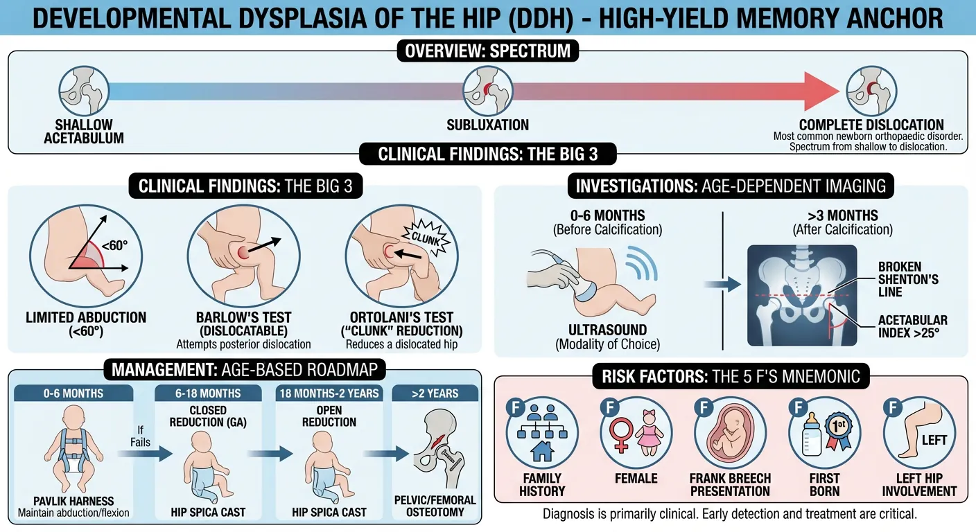
- Developmental Dysplasia of the Hip (DDH) is the most common orthopaedic disorder in newborns, encompassing a spectrum from a shallow acetabulum to complete dislocation [1].
- Diagnosis is primarily clinical, relying on specific physical examination maneuvers [1].
Key Clinical Findings
- Limited abduction of the flexed hip (<75°, or asymmetric) is a critical sign [1], [11].
- Barlow's test identifies dislocatable hips by attempting to dislocate the femoral head posteriorly [1].
- Ortolani's test reduces a dislocated hip, with a palpable 'clunk' indicating reduction [1].
Investigations
- Ultrasound is the imaging modality of choice in the first few months (before 4-6 months) due to uncalcified bone [1].
- X-rays are used from about 4 months, once the femoral head starts to ossify, with specific signs like an acetabular index >25° or a broken Shenton's line [1], [11].
Management by Age
- 0-6 months: Pavlik harness to maintain hip abduction and flexion [1].
- 6-18 months: Closed reduction under general anesthesia, followed by a hip spica cast, if Pavlik harness fails [1].
- 18 months-2 years: Open reduction with spica casting [1].
- >2 years: Pelvic and/or femoral osteotomy [1].
Risk Factors (5 Fs)
- Family history [1]
- Female [1]
- Frank breech presentation [1]
- First born [1], [3]
- LeFt hip involvement [1]
Definition / Overview
Developmental Dysplasia of the Hip (DDH) refers to an abnormal development of the hip joint. This can manifest as a shallow acetabulum (dysplasia), a partial displacement where articular surfaces maintain some contact (subluxation), or a complete separation of the joint (dislocation) [1]. It is recognized as the most common orthopaedic disorder observed in newborns [1].
Epidemiology
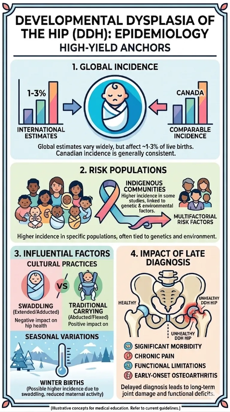
The incidence of DDH varies widely depending on diagnostic criteria, screening practices, and the population studied, but it is generally estimated to affect between 1 to 3% of live births [2, 3].
In Canada, studies have indicated a higher incidence in certain populations, for example, Indigenous communities, though the exact reasons are multifactorial and may include genetic predispositions and environmental factors [3].
There are known geographical and ethnic variations in incidence, with some populations exhibiting higher rates due to cultural practices, such as swaddling techniques that keep the hips extended and adducted, which can exacerbate or cause DDH [3, 4]. Conversely, cultures that traditionally carry infants with hips abducted and flexed tend to have lower rates [3]. Seasonal variation has also been observed: most studies report a higher incidence in winter births, possibly because infants are swaddled or dressed more tightly in colder months, although the mechanism is not established [3].
Despite advancements in screening and early detection, late diagnosis of DDH can lead to significant morbidity, characterized by pain, functional limitations, and early-onset osteoarthritis [1]. Therefore, understanding the epidemiology is crucial for targeted screening programs and effective prevention strategies.
Pathophysiology
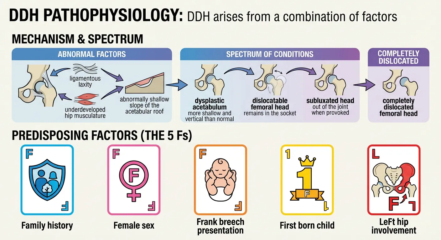
DDH arises from a combination of factors, including ligamentous laxity, underdeveloped hip musculature, and an abnormally shallow slope of the acetabular roof [1]. It presents as a spectrum of conditions, ranging in severity from a dysplastic acetabulum (more shallow and vertical than normal) to a completely dislocated femoral head [1]. Intermediate presentations include a dislocatable femoral head that remains in the socket, or a head that subluxates out of the joint when provoked [1].
Predisposing Factors (The 5 Fs)
Mnemonic: The 5 Fs that Predispose to DDH
- Family history [1]
- Female sex [1]
- Frank breech presentation [1]
- First born child [1]
- LeFt hip involvement [1]
Clinical Presentation
The diagnosis of Developmental Dysplasia of the Hip (DDH) is primarily clinical [1]. Key findings on physical examination include:
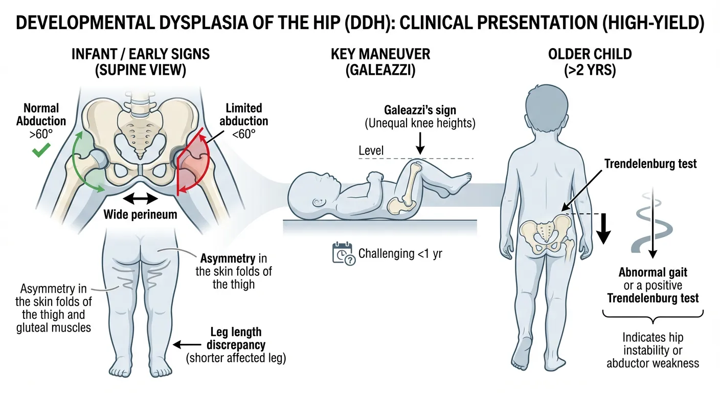
- Limited abduction: A crucial sign is reduced abduction of the flexed hip, typically less than 75 degrees, or clearly less than on the other side [1], [11].
- Leg length discrepancy: The affected leg may appear shorter, leading to asymmetry in the skin folds of the thigh and gluteal muscles [1].
- Wide perineum: A broader perineal space can be observed [1].
- Galeazzi's sign: When the hips and knees are flexed, the knees appear at unequal heights due to an apparent shorter femur on the affected side. This test can be challenging to perform accurately in children younger than one year [1].
- Trendelenburg test and gait: For older children (typically over 2 years of age), an abnormal Trendelenburg gait or a positive Trendelenburg test can indicate hip instability or abductor weakness [1].
Red Flags
Early identification of Developmental Dysplasia of the Hip (DDH) is critical, as diagnosis is primarily clinical [1]. Be vigilant for the following:
- Limited hip abduction: A reduced range of motion, specifically less than 75 degrees of abduction in a flexed hip or abduction that is asymmetric, is a significant red flag [1], [11].
- Asymmetry: Look for asymmetry in thigh or gluteal skin folds, or apparent leg length discrepancy [1].
- Positive Barlow's or Ortolani's tests: These maneuvers are definitive clinical indicators of hip instability in newborns [1].
- Predisposing factors (The 5 Fs): Patients with a history of family DDH, female sex, frank breech presentation, being first-born, or involvement of the left hip are at higher risk [1], [3].
⚠️ Do Not Miss: Limited abduction of the flexed hip is a primary indicator and should always prompt further investigation for DDH [1].
Diagnosis
The diagnosis of Developmental Dysplasia of the Hip (DDH) is fundamentally clinical, supported by imaging studies [1].
Physical Examination
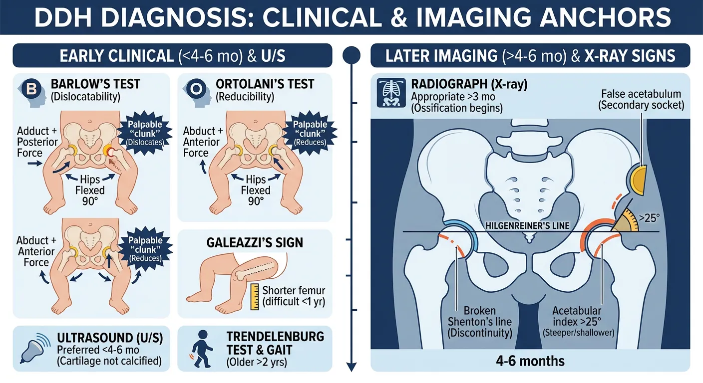
- Barlow's Test: This maneuver assesses for hip dislocatability [1]. The infant's hips and knees are flexed to 90 degrees, and the examiner grasps the thigh. The hips are fully adducted, and a posterior force is applied to attempt to dislocate the femoral head. A distinct palpable 'clunk' indicates a positive test, signifying that the hip can be dislocated [1].
- Ortolani's Test: This maneuver assesses for hip reducibility [1]. Starting from the same initial position as Barlow's, the examiner attempts to reduce a dislocated hip by gently abducting the hips while applying an anterior force with the fingertips. A palpable 'clunk' (not audible) indicates that a dislocated hip has been reduced [1].
- Galeazzi's Sign: Observed when the knees are at unequal heights with hips and knees flexed, appearing as a shorter femur on the affected side [1]. This test is difficult to interpret in children under one year of age [1].
- Trendelenburg Test and Gait: These are more useful for diagnosis in older children, typically over 2 years old [1].
Investigations
- Ultrasound (U/S): This is the preferred imaging modality during the first few months of life, as the infant's femoral head and acetabular cartilage are not yet calcified. Bony ossification typically begins around 4-6 months of age [1].
- Radiograph (X-ray): Radiographs become the main imaging study from about 4 to 6 months of age, once the femoral head has begun to ossify [1], [11].
X-ray Signs (typically seen at 4-6 months)
- False acetabulum: The development of a secondary socket due to chronic dislocation [1].
- Acetabular index >25°: Indicates a steeper and shallower acetabular roof [1].
- Broken Shenton's line: Discontinuity of the arc formed by the medial margin of the femoral neck and the superior margin of the obturator foramen [1].
- Femoral neck above Hilgenreiner's line: Hilgenreiner's line is a horizontal line drawn through the right and left triradiate cartilages. The femoral neck should normally lie below this line [1].
- Ossification centre outside of inner lower quadrant: Quadrants are formed by the intersection of Hilgenreiner's line and Perkin's line (a vertical line dropped from the lateral margin of the acetabulum). The femoral head ossification centre should typically be located in the inner lower quadrant [1].
Management
The management of Developmental Dysplasia of the Hip (DDH) is tailored to the child's age at diagnosis:
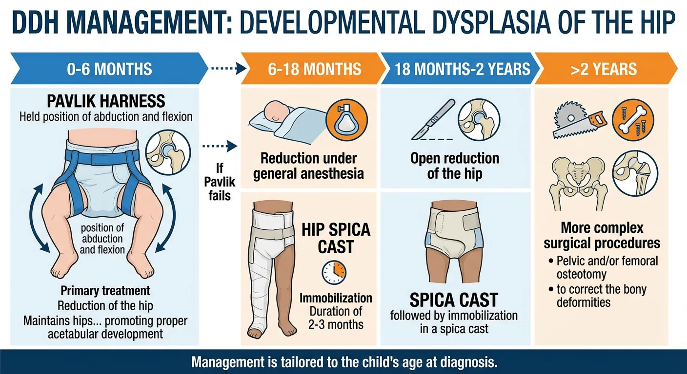
- 0-6 months: The primary treatment is reduction of the hip using a Pavlik harness. This device is designed to maintain the hips in a position of abduction and flexion, promoting proper acetabular development [1].
- 6-18 months: If a Pavlik harness fails or is initiated after 6 months of age, the next step involves reduction under general anesthesia, followed by immobilization in a hip spica cast, typically for about 12 weeks [1], [13].
- 18 months-2 years: At this age, open reduction of the hip is typically required, followed by immobilization in a spica cast [1].
- >2 years: For children over two years old, the treatment often involves more complex surgical procedures, such as a pelvic and/or femoral osteotomy, to correct the bony deformities [1].
Complications
Complications associated with Developmental Dysplasia of the Hip (DDH) can arise from both the condition itself and its treatment:
- Redislocation: The hip may dislocate again after initial reduction [1].
- Inadequate reduction: The hip may not be fully or stably reduced, leading to ongoing instability and potential long-term issues [1].
- Stiffness: Restricted range of motion in the hip joint can occur [1].
- Avascular Necrosis (AVN) of the femoral head: This is a significant complication that can occur at any stage of treatment [1]. It is primarily attributed to impingement of the medial circumflex femoral artery, often a result of severe and prolonged abduction and flexion positions maintained by devices like the Pavlik harness or spica cast [1].
Differential Diagnosis
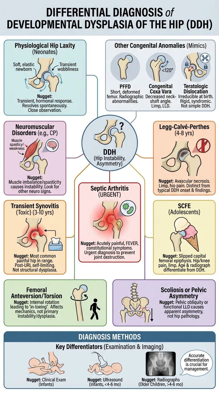
The differential diagnosis for developmental dysplasia of the hip (DDH) involves conditions that can present with similar clinical signs, particularly hip instability, asymmetry of gluteal folds, or limb length discrepancy in infants and young children [1]. Accurate differentiation is crucial for appropriate management and to prevent long-term complications.
Key conditions to consider include [1]:
- Physiological Hip Laxity: Many neonates, especially females, exhibit transient hip laxity that resolves spontaneously within the first few weeks of life. This is often an elastic response to maternal hormones and is typically not associated with true dysplasia. Close observation is usually sufficient, but persistent laxity warrants further investigation [1].
- Neuromuscular Disorders: Conditions such as cerebral palsy, spina bifida, and other muscular dystrophies can lead to hip instability, subluxation, or dislocation due to muscle imbalance or spasticity. These are usually distinguishable by the presence of other neurological signs and symptoms [5].
- Other Congenital Hip Anomalies: While DDH encompasses a spectrum, some other congenital anomalies can mimic its presentation. These include:
- Proximal Femoral Focal Deficiency (PFFD): A rare congenital anomaly characterized by a short, deformed femur, which can include hip instability or dislocation. Radiographs typically show characteristic femoral abnormalities [1].
- Congenital Coxa Vara: A deformity of the proximal femur where the neck-shaft angle is decreased. This can lead to a limp and limb length discrepancy but is distinct from DDH in its underlying pathology [1].
- Teratologic Dislocation: This refers to hip dislocations that are present at birth and irreducible, often associated with other severe congenital anomalies or syndromes (e.g., arthrogryposis, Larsen syndrome). The hip is fixed in a dislocated position, so the Barlow and Ortolani manoeuvres are negative while abduction is limited [11], [12].
- Transient Synovitis of the Hip (Toxic Synovitis): This is the most common cause of painful hip in children aged 3-10 years, presenting with limping and restricted hip movement. It is usually preceded by an upper respiratory infection and is self-limiting. It does not cause hip instability or structural dysplasia [6].
- Septic Arthritis of the Hip: A serious bacterial infection within the hip joint, presenting with an acutely painful hip, fever, and constitutional symptoms. It requires urgent diagnosis and treatment to prevent joint destruction. Pain and systemic illness are much more prominent than in DDH [7].
- Legg-Calvé-Perthes Disease: This condition involves avascular necrosis of the femoral head and typically affects children aged 4-8 years, presenting with a limp and hip pain. It is a later-onset condition than typical DDH and has distinct radiographic findings [8].
- Slipped Capital Femoral Epiphysis (SCFE): A condition where the head of the femur slips off the neck. It typically occurs in adolescents, often overweight males, presenting with hip or knee pain and a limp. The age of presentation and radiographic findings clearly differentiate it from DDH [9].
- Femoral Anteversion or Torsion: Common conditions where the femur is rotated internally, leading to an 'in-toeing' gait. While it can affect hip mechanics, it is not a primary hip instability or dysplasia [10].
Careful clinical examination, including Barlow and Ortolani maneuvers in infants, alongside appropriate imaging (ultrasound in infants, radiographs in older children), is essential to distinguish DDH from these other conditions [1, 2].
Exam Pearls
Performing a thorough hip examination is crucial for diagnosing DDH. Here are some key pearls:
Clinical Pearl: Diagnosis of DDH is clinical, making a meticulous physical exam paramount [1].
- Barlow's Test Execution: When performing Barlow's, remember to fully adduct the hips and apply a posterior force to attempt dislocation. The goal is to feel for a distinct 'clunk' as the hip dislocates [1].
- Ortolani's Test Execution: For Ortolani's, the movement is abduction while applying an anterior force. A palpable 'clunk' confirms that a dislocated hip has been reduced. Importantly, the 'clunk' should be felt, not heard, distinguishing it from benign clicks [1].
- Galeazzi's Sign Interpretation: While useful, Galeazzi's sign can be difficult to assess accurately in infants under one year of age due to their flexibility and limited cooperation [1].
- Trendelenburg for Older Children: The Trendelenburg test and assessment of gait are specifically useful for children over 2 years of age, as they can cooperate and bear weight [1].
- Limited Abduction: Always consider DDH if hip abduction is limited, especially if less than 75 degrees or asymmetric, as this is a consistent and important sign [1], [11].
Common Traps / Pitfalls
Common Exam Traps / Pitfalls in DDH Diagnosis
- Misinterpreting the 'Clunk': A common trap is confusing a benign click or snap (often heard) with the distinct, palpable 'clunk' of a dislocating or reducing hip. The key is that the DDH 'clunk' is felt, not just heard, as the femoral head moves in relation to the acetabulum [1].
- Difficulty with Galeazzi's Sign in Infants: Relying too heavily on Galeazzi's sign in children under one year can be misleading due to the inherent difficulty in assessing it accurately in very young, uncooperative infants [1].
- Over-Abduction with Pavlik Harness/Spica Cast: While necessary for treatment, excessive or prolonged forced abduction and flexion in a Pavlik harness or spica cast can lead to serious complications like avascular necrosis (AVN) of the femoral head due to impingement of the medial circumflex femoral artery. This highlights the importance of careful monitoring and correct application of these devices [1].
FAQ
Frequently Asked Questions about DDH
- Q: What is the most common orthopaedic disorder in newborns?
A: Developmental Dysplasia of the Hip (DDH) is the most common orthopaedic disorder in newborns [1]. - Q: When is ultrasound preferred over X-ray for DDH diagnosis?
A: Ultrasound is preferred in the first few months of life (typically up to 4-6 months) because the femoral head and acetabular cartilage are not yet calcified and are therefore not visible on X-ray [1]. X-rays become useful from about 4 months, once the femoral head begins to ossify [1], [11]. - Q: What are the '5 Fs' that predispose an infant to DDH?
A: The '5 Fs' are Family history, Female sex, Frank breech presentation, First born, and involvement of the LeFt hip [1], [3].
References
- [1] Nandhagopal T, Tiwari V, De Cicco FL. Developmental Dysplasia of the Hip. StatPearls [Internet], StatPearls Publishing; updated May 2024 (academic): https://www.ncbi.nlm.nih.gov/books/NBK563157/
- [2] Shaw BA, Segal LS; AAP Section on Orthopaedics. Evaluation and Referral for Developmental Dysplasia of the Hip in Infants. Pediatrics. 2016;138(6):e20163107 (guideline): https://doi.org/10.1542/peds.2016-3107
- [3] Loder RT, Skopelja EN. The Epidemiology and Demographics of Hip Dysplasia. ISRN Orthopedics. 2011;2011:238607 (academic): https://pmc.ncbi.nlm.nih.gov/articles/PMC4063216/
- [4] International Hip Dysplasia Institute. Hip-Healthy Swaddling (professional body): https://hipdysplasia.org/hip-healthy-swaddling/
- [5] OrthoInfo (AAOS). Cerebral Palsy (professional body): https://www.orthoinfo.org/diseases--conditions/cerebral-palsy/
- [6] Whitelaw CC, Varacallo MA. Transient Synovitis. StatPearls [Internet], StatPearls Publishing (academic): https://www.ncbi.nlm.nih.gov/books/NBK459181/
- [7] Vijayan V, Mabrouk A. Septic Arthritis of the Pediatric Hip. StatPearls [Internet], StatPearls Publishing (academic): https://www.ncbi.nlm.nih.gov/books/NBK459284/
- [8] OrthoInfo (AAOS). Perthes Disease (Legg-Calve-Perthes) (professional body): https://www.orthoinfo.org/diseases--conditions/perthes-disease/
- [9] OrthoInfo (AAOS). Slipped Capital Femoral Epiphysis (professional body): https://www.orthoinfo.org/diseases--conditions/slipped-capital-femoral-epiphysis-scfe/
- [10] OrthoInfo (AAOS). Intoeing (professional body): https://www.orthoinfo.org/diseases--conditions/intoeing/
- [11] Vaquero-Picado A, González-Morán G, Garay EG, Moraleda L. Developmental dysplasia of the hip: update of management. EFORT Open Rev. 2019;4(9):548-556 (academic): https://pmc.ncbi.nlm.nih.gov/articles/PMC6771078/
- [12] Noordin S, Umer M, Hafeez K, Nawaz H. Developmental dysplasia of the hip. Orthop Rev (Pavia). 2010;2(2):e19 (academic): https://pmc.ncbi.nlm.nih.gov/articles/PMC3143976/
- [13] de Witte PB, van Bergen CJA, de Geest BL, et al. Treatment of decentered developmental dysplasia of the hip under the age of 1 year: an evidence-based clinical practice guideline, Part 2. EFORT Open Rev. 2022;7(8):542-553 (academic): https://pmc.ncbi.nlm.nih.gov/articles/PMC9458947/
Further reading
- American Academy of Orthopaedic Surgeons. Detection and Nonoperative Management of Pediatric Developmental Dysplasia of the Hip in Infants up to Six Months of Age: Evidence-Based Clinical Practice Guideline (2014) (guideline): https://www.aaos.org/globalassets/quality-and-practice-resources/pddh/pediatric-developmental-dysplasia-hip-clinical-practice-guideline-4-23-19.pdf
- Patel H; Canadian Task Force on Preventive Health Care. Preventive health care, 2001 update: screening and management of developmental dysplasia of the hip in newborns. CMAJ. 2001;164(12):1669-1677 (guideline): https://pmc.ncbi.nlm.nih.gov/articles/PMC81153/
- OrthoInfo (AAOS). Developmental Dysplasia of the Hip (DDH) (professional body): https://www.orthoinfo.org/diseases--conditions/developmental-dislocation-dysplasia-of-the-hip-ddh/
- UpToDate. Developmental dysplasia of the hip: Clinical features and diagnosis (subscription) (academic): https://www.uptodate.com/contents/developmental-dysplasia-of-the-hip-clinical-features-and-diagnosis
- UpToDate. Developmental dysplasia of the hip: Epidemiology and pathogenesis (subscription) (academic): https://www.uptodate.com/contents/developmental-dysplasia-of-the-hip-epidemiology-and-pathogenesis
- Tönnis D. Congenital Dysplasia and Dislocation of the Hip in Children and Adults. Springer; 1987 (academic): https://doi.org/10.1007/978-3-642-71038-4