Supracondylar Humerus Fractures: A Clinical Reference
Comprehensive guide to pediatric supracondylar humerus fractures, covering epidemiology, classification, diagnosis, and management for medical students.
High-Yield Summary
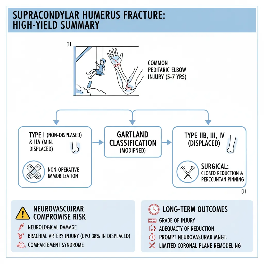
Supracondylar humerus fractures are common pediatric elbow injuries, accounting for up to 18% of all pediatric fractures and 60% of elbow fractures, typically occurring in children aged 5-7 years due to falls on an outstretched arm [1]. These fractures are classified using the modified Gartland system, guiding treatment from non-operative immobilization for non-displaced (Type I) and minimally displaced (Type IIA) fractures, to surgical closed reduction and percutaneous pinning for displaced (Type IIB, III, IV) injuries [1]. A critical aspect of management is a thorough neurovascular assessment, as these injuries carry significant risks of neurological damage, vascular injury, and compartment syndrome, with brachial artery injury reported in up to 38% of displaced cases [1]. Long-term outcomes are influenced by factors like the grade of injury, adequacy of reduction, and prompt management of neurovascular compromise, although remodeling potential for coronal plane deformities is limited [1].
Definition / Overview
Supracondylar humerus fractures involve the region of the distal humerus just above the elbow joint. These fractures are notably more prevalent in the pediatric population compared to adults, often resulting from children using an outstretched arm to protect themselves during a fall [1]. They represent a substantial portion of pediatric fractures, constituting up to 18% of all pediatric fractures and 60% of elbow fractures in children [1]. Despite advancements in evaluation and treatment, these injuries continue to present considerable challenges to orthopedic surgeons [1].
Epidemiology
- Prevalence: The supracondylar region of the distal humerus is the most frequently fractured area around the elbow in children [1].
- Age Group: Most commonly occurs in children between 5 and 7 years old, with a median age of approximately six years. The incidence gradually declines with increasing age [1].
- Gender: Some studies indicate a higher prevalence in males, others in females, while a recent epidemiological study found no significant gender difference [1].
- Fracture Type: Extension-type fractures are considerably more common than flexion-type, typically caused by a fall on an outstretched hand, and account for up to 95% of supracondylar fractures [1]. Flexion-type fractures are more prevalent in older children [1].
- Associated Factors: Ligamentous laxity in children contributes to the occurrence of extension-type fractures by allowing hyperextension of the elbow [1]. These fractures are reported to be more frequent on weekends and during summer months [1].
- Laterality: Often occurs in the non-dominant upper extremity [1].
- Open Fractures: The incidence of open supracondylar fractures is around 1% [1].
Pathophysiology
The distal humerus has a unique anatomical structure that makes the supracondylar region prone to fracture [1].
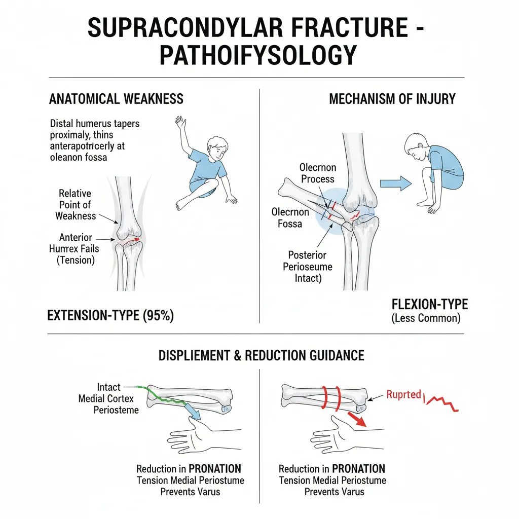
- Anatomical Weakness: The distal humerus tapers proximally from the metaphysis to the diaphysis, and thins anteroposteriorly where the medial and lateral columns merge at the olecranon fossa. This creates a relative point of weakness in the supracondylar area [1].
- Mechanism of Injury (Extension-Type): Most fractures (up to 95%) are extension-type, occurring when a child falls on an outstretched arm, causing hyperextension of the elbow [1]. This forces the olecranon process to engage the olecranon fossa, generating significant stress on the distal humerus [1]. The anterior humeral cortex fails under tension, leading to fracture, while the posterior cortex and periosteum may remain intact [1].
- Mechanism of Injury (Flexion-Type): Less common, these fractures occur with a fall directly onto a flexed elbow [1].
- Displacement and Reduction Guidance: The direction of the distal fragment's displacement informs the reduction maneuver [1].
- Posteromedial displacement: Often involves an intact medial periosteum. Reduction is performed in pronation to tension the medial periosteum and prevent varus malalignment [1].
- Posterolateral displacement: Tends to rupture the medial periosteum. Reduction is performed in supination [1].
Clinical Presentation
History
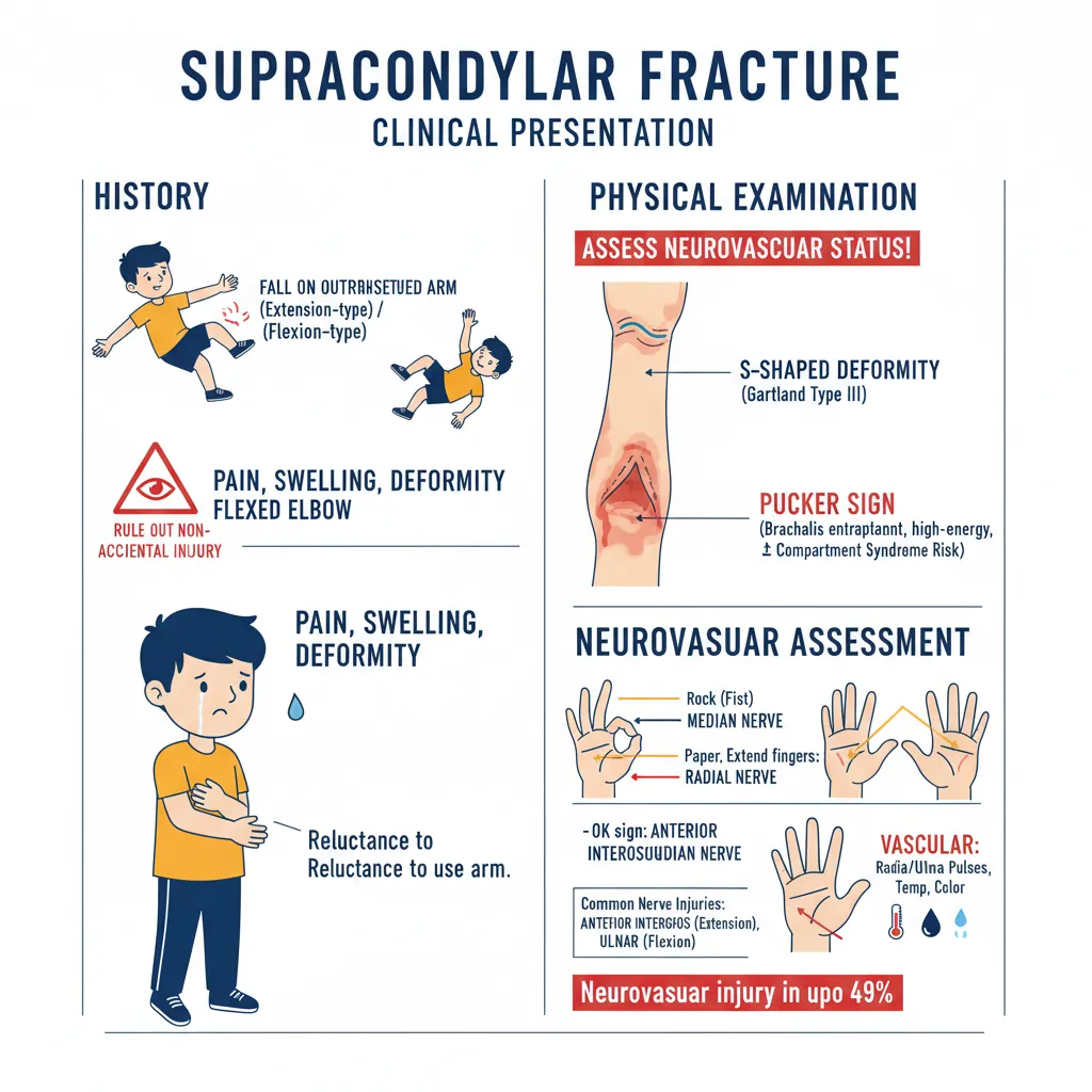
- A common history is a fall on an outstretched arm (for extension-type fractures) or, less commonly, a fall on a flexed elbow [1].
- It is crucial to consider and rule out non-accidental injury, though this accounts for less than 0.5% of cases [1].
- Patients or carers typically report pain, swelling, or deformity of the affected limb [1].
- Young children may be reluctant to use the injured arm [1].
Physical Examination
A comprehensive assessment of the soft tissue integrity and distal neurovascular status is mandatory, along with evaluation of any other associated injuries [1].
- Appearance: The limb may present with swelling, bruising, and deformity. Any bleeding could indicate an open fracture [1].
- S-shaped Deformity: A classic S-shaped deformity is typical for 'off-ended' Gartland Type III injuries [1].
- 'Pucker Sign': An indentation in the antecubital fossa may signify that the proximal fragment has 'button-holed' through the brachialis muscle. This is a red flag for high-energy injury and potential brachial artery or median nerve damage, as these structures can become entrapped during reduction [1]. Patients with a pucker sign are also at increased risk for compartment syndrome [1].
- Neurovascular Assessment: This is paramount and must be meticulously documented and repeated after any intervention [1].
- Nerves: Assess motor and sensory functions of the median, anterior interosseous, ulnar, and radial nerves [1]. Neurovascular injuries have been reported as high as 49% [1].
- Common Nerve Injuries: Anterior interosseous nerve in extension-type fractures, and ulnar nerve in flexion-type injuries [1].
- Pediatric Nerve Assessment: Use 'rock, paper, scissors, OK' games:
- Rock (fist): Evaluates median nerve [1].
- Paper (extend fingers): Assesses radial nerve [1].
- Scissors (abduct fingers): Checks ulnar nerve [1].
- OK sign: Evaluates anterior interosseous nerve [1].
- Vascular Status: Examine distal limb perfusion, including radial and ulnar pulses, temperature, color of the ipsilateral hand, and capillary refill time [1]. Vascular compromise is reported in 10-20% of displaced supracondylar fractures [1].
- 'Pulseless Pink, Perfused Hand': Indicates good collateral circulation. Requires urgent management [1].
- 'Pulseless, Pale, Cold Hand': Evidences an ischemic limb, constituting a surgical emergency [1].
- Combined Deficit: The presence of both neurological deficit and a pulseless hand strongly predicts vascular compromise with entrapment of both artery and nerve [1]. Brachial artery injury can occur in up to 38% of displaced Gartland III fractures [1].
- 'Pulseless, Pale, Cold Hand': This is an immediate surgical emergency indicating limb ischemia [1].
- Neurological Deficit + Pulseless Hand: Highly predictive of vascular compromise with nerve and artery entrapment [1].
- Brachial Artery Injury: Can occur in up to 38% of displaced Gartland Type III fractures [1].
- Traumatic Neuropraxia: Incidence up to 11.3%, with the anterior interosseous nerve most commonly affected in extension-type, and ulnar nerve in flexion-type injuries [1].
- 'Pucker Sign': An indentation in the antecubital fossa indicates a high-energy injury and significant risk of brachial artery or median nerve entrapment and compartment syndrome [1].
- Open Fracture: Any bleeding around the elbow points to an open fracture, requiring urgent surgical attention [1].
- Non-Accidental Injury (NAI): Although rare (<0.5%), NAI must always be considered and excluded in pediatric fractures [1].
- Medial Column Comminution in Gartland II: Can lead to medial column collapse and cubitus varus deformity if not recognized and managed appropriately [1].
- Anteroposterior (AP) Radiographs:
- A transverse or short oblique fracture line may be visible in the supracondylar region; it can be subtle or missed in non-displaced fractures [1].
- Baumann's Angle (Humerocapitellar Angle): The angle between the longitudinal axis of the humerus and a line tangential to the capitellar physis. Normal range is 64-81 degrees [1]. An increased angle suggests cubitus varus deformity and is a reliable guide for coronal plane alignment [1].
- Radiocapitellar Line: A line through the radial long axis should always intersect the capitellum. Disruption suggests an associated radial head/neck fracture [1].
- Lateral Radiographs:
- Posterior Fat Pad Sign: An elevated posterior fat pad indicates an intra-articular effusion, often signifying an occult or non-displaced intra-articular fracture within the elbow [1].
Clinical Pearl: A positive posterior fat pad sign in a child with elbow trauma should prompt consultation with an orthopedic surgeon, as it suggests an intra-articular fracture even if not immediately visible [1].
- Anterior Humeral Line: A line drawn along the anterior humeral cortex on the lateral view should bisect the middle third of the capitellum (or anterior one-third in children under four years) [1]. If this line passes anterior to the capitellum, it indicates an extension-type fracture [1].
- Type I: Non-displaced or minimally displaced (<2 mm) fractures [1].
- Type IIA: Displaced fractures (>2 mm) with an intact posterior hinge (cortex) [1].
- Type IIB: Displaced fractures with an intact posterior hinge and associated malrotation [1].
- Type III: Completely displaced fractures with disruption of the posterior periosteum [1].
- Type IV: Completely displaced fractures with multidirectional instability, identified intra-operatively due to complete periosteal tear [1].
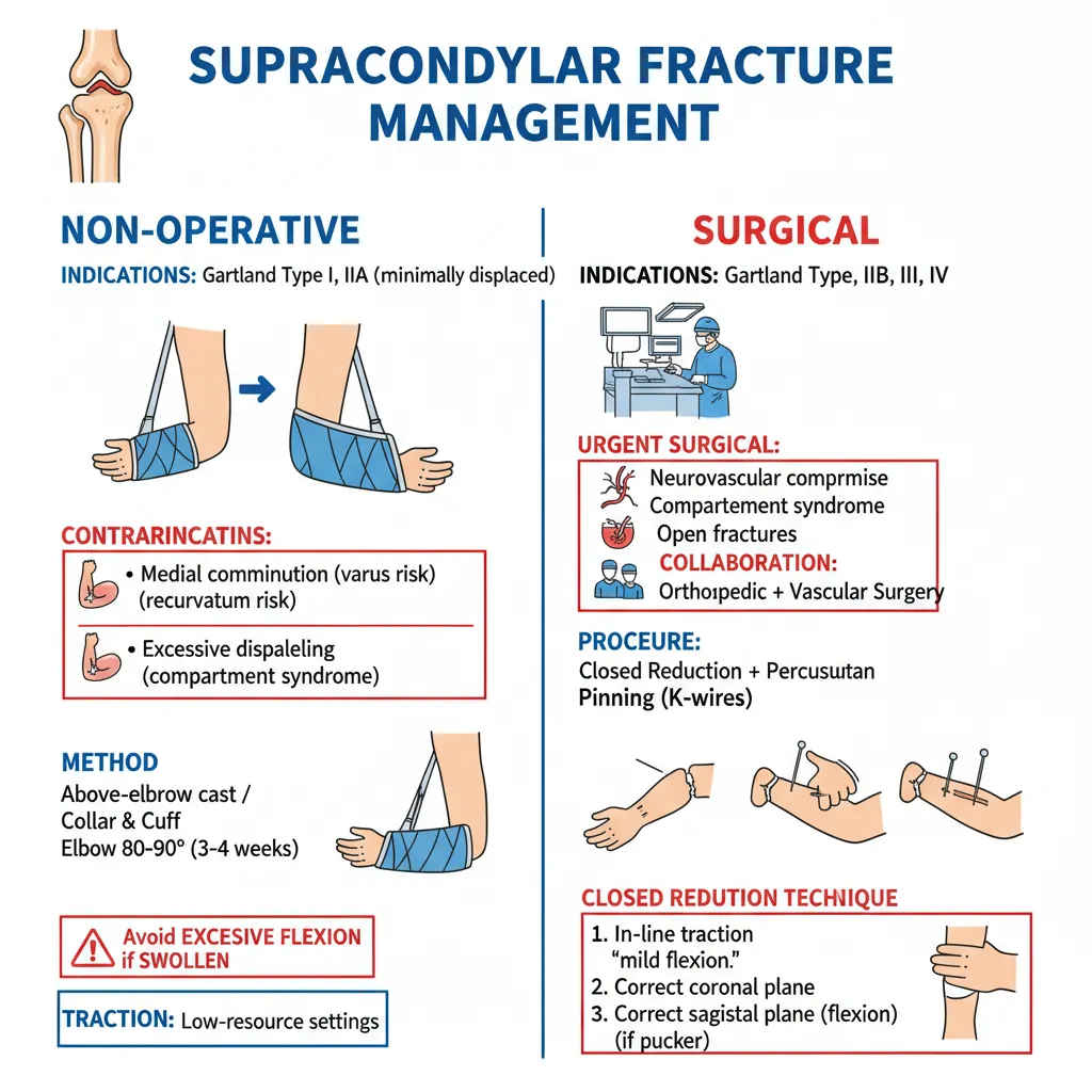
- Indications: Acceptable for non-displaced Gartland Type I and minimally displaced Gartland Type IIA fractures [1].
- Contraindications: Medial column comminution (risk of cubitus varus), posterior displacement (risk of recurvatum if anterior humeral line doesn't bisect capitellum), and excessive swelling (risk of compartment syndrome) contraindicate non-operative management for Gartland Type II fractures [1].
- Method: Immobilization in a collar and cuff or an above-elbow cast with the elbow flexed 80-90 degrees for 3-4 weeks [1].
- Considerations: Avoid excessive flexion in patients with significant elbow swelling to prevent compartment syndrome [1]. Above-elbow casts offer better pain relief for Gartland I fractures than a collar and cuff [1].
- Traction: Historically used for displaced fractures and may still be employed in low-resource settings [1].
- Indications: Displaced Gartland Type II (IIB), Type III, and Type IV fractures [1].
- Urgent Surgical Indications: Neurovascular compromise, compartment syndrome, and open fractures [1].
- Procedure: Typically involves closed reduction and percutaneous pinning with K-wires for closed injuries [1].
- Collaboration: Orthopedic surgeons should be prepared to manage associated neurovascular injuries and have vascular surgical assistance available if needed [1].
- Indications: Gartland IIB and III fractures. Also for Gartland IIA if acceptable reduction isn't achieved non-operatively or if contraindications to non-operative treatment exist [1].
- Closed Reduction Technique:
- In-line traction with 'milking' of soft tissues (if pucker sign present) to achieve length and release soft tissues [1]. Traction is applied with the elbow in slight flexion [1].
- Correction of coronal plane displacement (medial/lateral translation, varus/valgus angulation) [1].
- Correction of rotational deformity: Pronation for posteromedial displacement (internal rotation) to tension the medial periosteum; supination for posterolateral (external) rotation [1].
- Correction of sagittal plane deformity by hyperflexion for extension-type fractures (or extension for flexion-type) [1].
- The 'joystick technique' is used for multi-directionally unstable fractures [1].
- Percutaneous Pinning Technique:
- Pin Configurations: Lateral pins (two or three) or crossed pins (medial and lateral) bicortical constructs [1].
- Lateral Pins: Preferred to reduce iatrogenic ulnar nerve injury risk [1]. Inserted in parallel or divergent manner with maximal separation; divergent pattern is more stable [1]. A capitellar starting point and trans-olecranon fossa four-cortex purchase can enhance rigidity [1]. 2mm pins offer better biomechanical strength than 1.6mm pins [1].
- Crossed Pins: Improve torsional rigidity but increase ulnar nerve injury risk [1]. A medial incision is essential to protect the ulnar nerve before medial pin insertion [1]. Extending the elbow during medial pin insertion displaces the nerve posteriorly, further reducing risk [1]. Avoid percutaneous medial pin insertion without direct visualization of the nerve [1].
- Indications: When closed reduction fails (e.g., due to trapped soft tissue or neurovascular structures) or for open fractures [1].
- Vascular Exploration: Required if perfusion is not restored post-reduction and fixation [1].
- Approach: An anterior approach is recommended [1]. Medial, lateral, and posterior approaches are alternatives [1].
- Urgent Indication: Patients with a 'pulseless, pale hand' require immediate vascular exploration by an experienced surgeon or in conjunction with a vascular surgeon [1]. Initial attempt at closed reduction and fixation should be made to see if perfusion is restored [1].
- 'Pink, Perfused, Pulseless Hand' Post-Reduction: Management is controversial [1]. Close observation is needed, as some may still develop ischemia requiring urgent vascular exploration [1]. Non-operative management with close monitoring can yield acceptable results [1].
- Percutaneous pins are typically removed 3-4 weeks post-operatively, after which children can begin mobilization [1].
- Initial stiffness is common but improves rapidly, often achieving 98% of normal range of motion within 12 months [1]. Older children may regain motion more slowly [1].
- Physiotherapy has not shown to improve outcomes in randomized controlled trials [1].
- Neurological Injury:
- Approximately 11.3% incidence of traumatic neuropraxia [1].
- Anterior Interosseous Nerve Palsy: Most common in extension-type fractures [1].
- Median Nerve Palsy: Can be traumatic or iatrogenic [1].
- Radial Nerve Palsy: More common with posteromedial displacement [1].
- Ulnar Nerve Palsy: Most common in flexion-type fractures and a risk of medial pin placement [1].
- Vascular Injury:
- Brachial artery injury is more common in completely displaced Gartland Type III (and IV) injuries [1].
- Compartment Syndrome:
- Can develop in the forearm due to impaired perfusion (brachial artery injury) and inadequate collateral circulation [1].
- Volkmann Ischemic Contracture:
- A relatively rare but severe complication resulting from forearm compartment syndrome and delayed treatment of arterial injury leading to forearm ischemia [1].
- Malunion and Deformity:
- Can occur in sagittal, coronal, and axial planes [1].
- Recurvatum Deformity: Sagittal plane deformity. Younger children have better remodeling potential for sagittal plane deformities [1].
- Cubitus Varus (Gunstock Deformity): Coronal and axial plane deformities have minimal remodeling potential [1]. Can result from medial column comminution leading to collapse, or residual internal rotation [1].
- Cubitus Valgus: Can occur from inadequate reduction and malunion, potentially leading to tardy ulnar nerve palsy [1].
- Stiffness:
- Range of movement improves rapidly post-surgery, reaching up to 98% of normal within 12 months [1]. Stiffness is more commonly associated with open reduction and malunion [1].
- Pin-site Infection: A risk with percutaneous pinning [1].
- Pin-site Migration: A risk with percutaneous pinning [1].
- Radial head fractures [1]
- Distal humeral medial or lateral condyle fractures [1]
- Transphyseal fractures of the distal humerus (non-accidental injury should be excluded in these cases) [1]
- Pulled elbow (usually diagnosed by history, radiographs often not needed) [1]
- [1] Hope N, Varacallo MA. Supracondylar Humerus Fractures. StatPearls [Internet], StatPearls Publishing (academic): https://www.ncbi.nlm.nih.gov/books/NBK560933/
Red Flags
Red Flags
⚠️ Do Not Miss: Neurovascular Compromise:
⚠️ Do Not Miss: Compartment Syndrome: Patients are at risk of developing compartment syndrome, particularly with significant elbow swelling or compromised vascularity [1]. Increasing analgesia requirements can be a subtle sign [1].
Diagnosis
Radiographic Evaluation
Plain anteroposterior (AP) and lateral radiographs of the elbow are essential for diagnosis, determining fracture type (extension vs. flexion), displacement, malrotation, and comminution [1]. Interpreting pediatric elbow radiographs requires knowledge of the predictable ossification centers [1].
Gartland Classification of Supracondylar Humerus Fractures
The modified Gartland classification is widely used to describe fracture patterns and guide treatment [1]. Fractures are categorized as extension or flexion-type based on the mechanism and distal fragment displacement. Extension-type are most common, with the distal fragment extended relative to the proximal [1].
Angiography
Angiography is generally not recommended before reduction in cases of vascular compromise, as it delays critical treatment [1]. The Pediatric Orthopedic Society of North America (POSNA) supports this guideline [1].
Management
All patients should initially be assessed following Advanced Trauma Life Support (ATLS) principles, and non-accidental injury must be excluded in pediatric cases [1].
Non-Operative Management
Surgical Management
Closed Reduction and Percutaneous Pinning
Open Reduction and Percutaneous Pinning
Vascular Exploration
Postoperative and Rehabilitation Care
Complications
Supracondylar humerus fractures, if not managed appropriately, can lead to various complications [1].
Differential Diagnosis
Given the largely cartilaginous nature of the pediatric elbow, interpreting radiographs can be challenging, especially in very young children [1]. Differential diagnoses for elbow trauma in children include:
Exam Pearls
Clinical Pearl: The presence of a 'pucker sign' on clinical examination strongly indicates a severe soft tissue injury and a high risk of neurovascular compromise [1].
Clinical Pearl: Refer patients with a positive posterior fat pad sign to an orthopedic surgeon for further review, as it signifies an intra-articular fracture, even if not overtly visible [1].
Clinical Pearl: Intra-operatively, in addition to standard AP and lateral views, consider obtaining internal oblique and external oblique views to improve visualization of the medial and lateral columns for reduction assessment [1].
Clinical Pearl: When performing a neurological exam on children, use interactive methods like 'rock, paper, scissors, OK' to assess median, radial, ulnar, and anterior interosseous nerve function respectively [1].
Common Traps / Pitfalls
Common Exam Trap: Failing to recognize the subtle signs of neurovascular compromise, particularly a 'pulseless pink, perfused hand,' which still requires urgent management and close observation despite good perfusion [1]. A delay in recognizing evolving ischemia can lead to severe complications like Volkmann ischemic contracture [1].
Common Pitfall: Pin placement during percutaneous pinning must ensure maximal engagement at the fracture site. Lateral pins that cross at the fracture site or crossed pins that cross only in the distal fragment offer little to no rotational stability, increasing the risk of unstable fixation [1].
Common Pitfall: Not appreciating the limited remodeling potential of the distal humerus, especially for coronal plane deformities like cubitus varus, which will not remodel significantly, even in younger children [1]. This underscores the importance of achieving anatomical reduction [1].
Common Pitfall: Neglecting to make a protective medial incision to shield the ulnar nerve when inserting a medial pin in a crossed-pin construct, which significantly increases the risk of iatrogenic ulnar nerve injury [1].