
Hyponatremia USMLE: Diagnosis, Causes & Treatment
Master hyponatremia with this comprehensive USMLE guide covering diagnosis, causes, SIADH, treatment, complications, exam pearls, and management strategies.
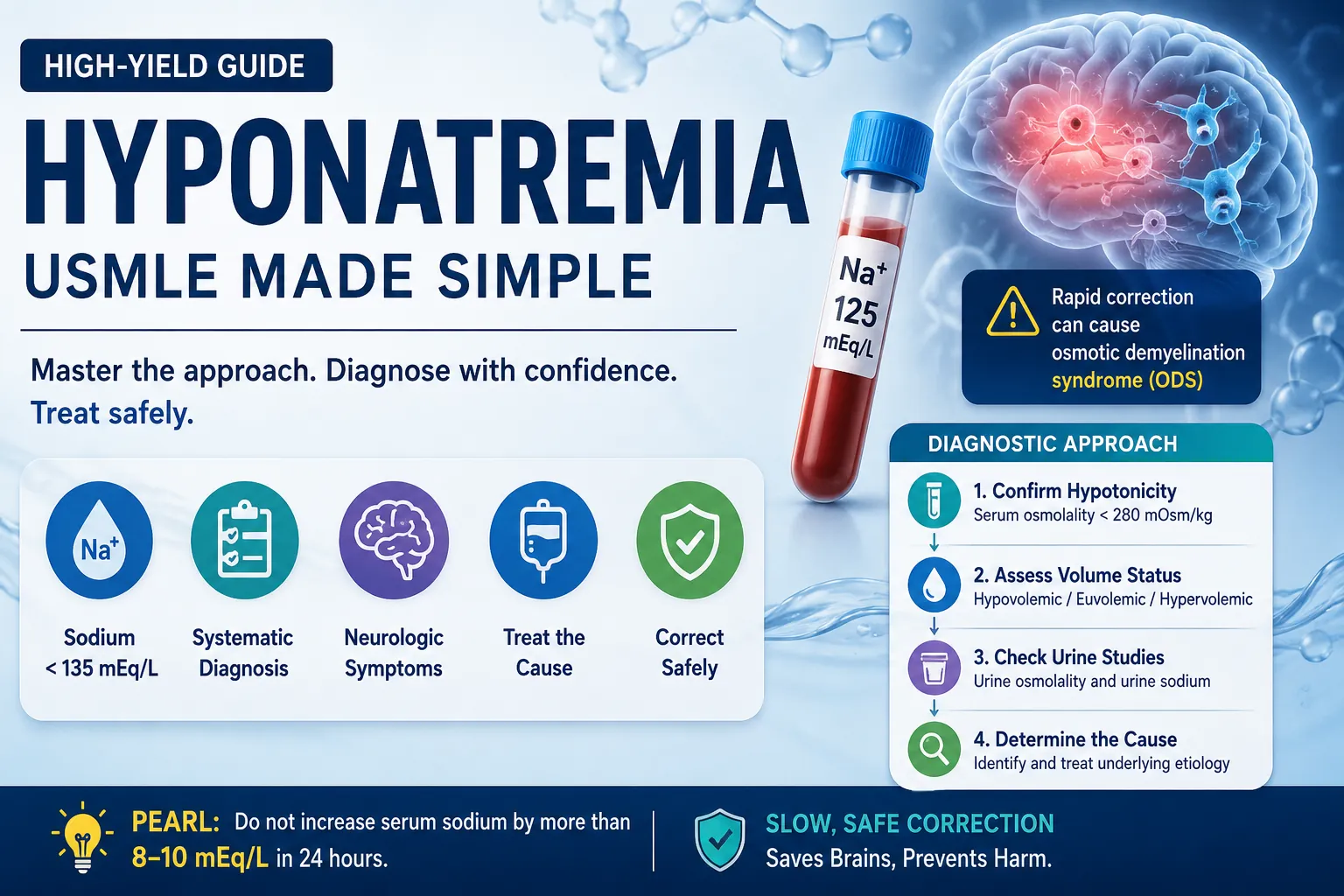
Hyponatremia is one of the most frequently tested electrolyte disorders in USMLE Step 1 and Step 2 CK. Questions on this topic appear in internal medicine, nephrology, endocrinology, neurology, emergency medicine, and critical care. Because of its wide clinical relevance, understanding hyponatremia is essential for success on board examinations and in clinical practice.
Many students struggle with Hyponatremia USMLE questions because they rely on memorizing causes instead of following a structured diagnostic approach. Fortunately, a simple framework can make even complex clinical scenarios easier to solve.
This guide explains everything you need to know about hyponatremia, including its pathophysiology, classification, causes, diagnosis, management, complications, and the high-yield concepts most commonly tested on the USMLE. You'll also find clinical pearls, exam tips, and common pitfalls to help strengthen your clinical reasoning.
If you're preparing for your next board exam, explore more evidence-based learning resources on the Sentia Blog at www.mysentia.app/blogs. Sentia also provides additional study materials designed to help medical students build lasting clinical knowledge through structured, high-yield learning.
High-Yield Summary
Before diving into the details, review these essential facts. They highlight the concepts most frequently tested in Hyponatremia USMLE questions.
| Topic | High-Yield Fact |
|---|---|
| Definition | Serum sodium below 135 mEq/L.[1] |
| Most Common Cause | SIADH is the leading cause in hospitalized patients.[2] |
| Most Important Diagnostic Step | Confirm serum osmolality before assessing volume status.[3] |
| Most Dangerous Acute Complication | Cerebral oedema caused by rapid water movement into brain cells.[4] |
| Most Dangerous Treatment Complication | Osmotic Demyelination Syndrome from correcting sodium too quickly.[4] |
| Safe Correction Rate | No more than 8-10 mEq/L in 24 hours.[4][5] |
| USMLE Tip | Determine osmolality first, volume status second, then interpret urine studies. |
Clinical Pearl: In most patients, hyponatremia results from excess body water rather than sodium deficiency. This principle explains why fluid restriction often plays a key role in treatment.
Learning Objectives
After studying this guide, you should be able to:
- Define hyponatremia and explain why it develops.
- Classify hyponatremia using serum osmolality and volume status.
- Apply a structured diagnostic approach to patients with low serum sodium.
- Differentiate SIADH from other common causes of hyponatremia.
- Select the appropriate treatment based on symptom severity and underlying cause.
- Recognize common USMLE pitfalls and avoid management errors.
What Is Hyponatremia?
Hyponatremia occurs when the serum sodium concentration falls below 135 mEq/L.[1] It is the most common electrolyte disorder encountered in clinical practice and one of the highest-yield topics tested on medical licensing examinations.
Although laboratory reports describe hyponatremia as a low sodium level, the disorder usually reflects an imbalance between sodium and water rather than an absolute sodium deficiency. In many patients, excess water dilutes the sodium concentration, resulting in a lower measured serum sodium level.
As the sodium concentration falls, water moves into cells by osmosis. If the drop occurs rapidly, brain cells begin to swell. This swelling increases intracranial pressure and may lead to headache, confusion, seizures, coma, or even death if left untreated.[3][4]
Symptoms Depend on Two Things
The symptoms of hyponatremia depend on two things:
- How low the sodium level becomes
- How quickly the sodium level falls
A person with chronic hyponatremia may have a sodium level of 120 mEq/L and only mild symptoms. The brain has enough time to adjust to the gradual change.
A rapid drop is much more dangerous. For example, if the sodium level falls from 140 mEq/L to 125 mEq/L within a few hours, brain swelling can occur. This may lead to confusion, seizures, or even coma.
This concept appears frequently in Hyponatremia USMLE questions. Before deciding on the diagnosis or treatment, always ask how low the sodium is and how fast it dropped.
USMLE Tip: Always ask two questions before choosing treatment:
- Is the hyponatremia acute or chronic?
- Is the patient symptomatic or asymptomatic?
The answers determine both the urgency and the type of treatment required.
Understanding Sodium Homeostasis
To understand hyponatremia, you must first understand how the body regulates sodium and water.
Sodium is the main positively charged ion in the extracellular fluid. It plays a central role in maintaining serum osmolality, regulating fluid balance, supporting nerve conduction, and preserving normal muscle function.[5]
The body maintains serum sodium within a narrow range through the coordinated actions of three major regulatory systems:
- Antidiuretic hormone (ADH)
- The renin-angiotensin-aldosterone system (RAAS)
- The thirst mechanism
Together, these systems continuously adjust water intake and water excretion to keep serum osmolality stable.
Antidiuretic Hormone (ADH)
ADH, also known as vasopressin, is produced in the hypothalamus and released from the posterior pituitary gland. Two main signals stimulate its release:
- Increased serum osmolality
- Reduced effective circulating blood volume
After release, ADH acts on the collecting ducts of the kidneys, increasing water reabsorption. As more water returns to the bloodstream, the kidneys produce a smaller volume of concentrated urine.
This response helps preserve blood pressure and restore normal osmolality. However, excessive ADH activity can also dilute serum sodium and contribute to hyponatremia.
The Renin-Angiotensin-Aldosterone System (RAAS)
The RAAS helps maintain blood pressure and sodium balance.
When blood pressure falls or kidney perfusion decreases, the kidneys release renin. This activates a hormonal cascade that produces angiotensin II and stimulates aldosterone secretion.
Aldosterone increases sodium reabsorption in the distal nephron. Water follows sodium, helping restore circulating blood volume.
The Thirst Mechanism
The hypothalamus also regulates thirst. As serum osmolality rises, thirst increases, encouraging water intake.
Normally, these three systems work together to maintain fluid balance. However, diseases that disrupt this regulation can lead to dilutional hyponatremia.
Clinical Pearl: Most Hyponatremia USMLE questions focus on abnormalities in water regulation rather than problems with sodium intake alone.
Continue Learning
Want to strengthen your understanding of renal physiology before moving on? Explore more USMLE-focused learning resources on the Sentia Blog:
- Blog: www.mysentia.app/blogs
- About Sentia: www.mysentia.app/home
- Home: www.mysentia.app/home
You can also review the National Institute of Diabetes and Digestive and Kidney Diseases (NIDDK) overview of electrolyte disorders for additional background reading: https://www.niddk.nih.gov/
Classification of Hyponatremia USMLE
One of the easiest ways to answer Hyponatremia USMLE questions is to classify the disorder correctly before searching for the cause. Most board-style questions follow the same sequence:
- Determine the serum osmolality.
- Assess the patient's volume status.
- Identify the underlying cause.
Following this framework narrows the differential diagnosis and points you toward the correct treatment.
Step 1: Classify by Serum Osmolality
Serum osmolality helps determine whether the low sodium level reflects true hyponatremia or another process.
| Type | Serum Osmolality | Common Causes |
|---|---|---|
| Hypotonic Hyponatremia | Less than 280 mOsm/kg | SIADH, heart failure, cirrhosis, diuretics |
| Isotonic Hyponatremia | 280-295 mOsm/kg | Severe hyperlipidaemia, hyperproteinaemia (pseudohyponatremia) |
| Hypertonic Hyponatremia | Greater than 295 mOsm/kg | Hyperglycaemia, mannitol therapy |
Hypotonic Hyponatremia
Hypotonic hyponatremia is the most common and most clinically significant form of hyponatremia. It accounts for the majority of cases seen in hospitals and appears most frequently on USMLE examinations.[3]
In this condition, excess water lowers both serum sodium and serum osmolality. Because water moves into cells, patients are at risk of cerebral oedema and neurological complications.
Isotonic Hyponatremia
Isotonic hyponatremia, also known as pseudohyponatremia, does not reflect a true decrease in serum sodium.
Instead, laboratory measurement errors occur when extremely high lipid or protein levels reduce the proportion of plasma occupied by water. Modern laboratory methods have made this condition less common, but it remains a favorite USMLE distractor.
Exam Trap: Pseudohyponatremia does not require treatment with saline because the patient's serum osmolality remains normal.
Hypertonic Hyponatremia
Hypertonic hyponatremia develops when another osmotically active substance draws water from the intracellular space into the bloodstream.
Common causes include:
- Severe hyperglycaemia
- Mannitol administration
Although the measured sodium concentration decreases, the underlying problem is excess extracellular osmoles rather than sodium deficiency.
USMLE Tip: Whenever you see severe hyperglycaemia, calculate the corrected serum sodium before diagnosing true hyponatremia.
Step 2: Classify by Volume Status
Once you've confirmed hypotonic hyponatremia, determine the patient's extracellular fluid volume.
This step identifies the underlying mechanism and guides treatment.
| Volume Status | What Happens? | Common Causes |
|---|---|---|
| Hypovolaemic | Sodium and water decrease, but sodium loss exceeds water loss. | Vomiting, diarrhoea, burns, thiazide diuretics, adrenal insufficiency |
| Euvolaemic | Water increases slightly while total body sodium remains relatively unchanged. | SIADH, hypothyroidism, adrenal insufficiency, primary polydipsia |
| Hypervolaemic | Sodium and water both increase, but water retention exceeds sodium retention. | Heart failure, cirrhosis, nephrotic syndrome, chronic kidney disease |
Remember this sequence for every Hyponatremia USMLE question:
Osmolality → Volume Status → Urine Studies → Diagnosis
Pathophysiology
Understanding the pathophysiology of hyponatremia makes diagnosis much easier. Instead of memorizing dozens of causes, focus on why excess water accumulates.
In most cases, the kidneys retain more water than the body can safely eliminate. As water increases, the serum sodium concentration falls.
Three main mechanisms explain this process.
1. Excess Antidiuretic Hormone (ADH)
Excessive ADH activity is the most common mechanism behind hyponatremia.
ADH increases water reabsorption in the collecting ducts of the kidneys. As a result, water returns to the bloodstream without adding sodium. Consequently, serum sodium becomes diluted.
This mechanism explains Syndrome of Inappropriate Antidiuretic Hormone Secretion (SIADH), which remains the leading cause of euvolaemic hyponatremia.[2]
2. Excess Water Intake
Some patients drink more water than their kidneys can excrete.
This occurs in conditions such as:
- Primary (psychogenic) polydipsia
- Beer potomania
- Low-solute diets
When water intake exceeds renal excretory capacity, serum sodium gradually falls.
3. Reduced Water Excretion
Several diseases reduce the kidneys' ability to eliminate free water.
These include:
- Heart failure
- Cirrhosis
- Nephrotic syndrome
- Advanced chronic kidney disease
Although these patients often retain sodium, they retain even more water, resulting in dilutional hyponatremia.
Why Brain Cells Swell
Water always moves toward the area with the higher concentration of dissolved particles.
When serum sodium falls, the fluid outside brain cells becomes less concentrated than the fluid inside them. Water then moves into the brain cells through osmosis.
If this process happens quickly, brain cells swell. The resulting increase in intracranial pressure produces symptoms such as headache, confusion, seizures, and coma.[4]
Patients with chronic hyponatremia adapt over time by reducing intracellular osmolytes. This adaptation explains why chronic hyponatremia often causes fewer symptoms than acute hyponatremia.
Clinical Pearl: The speed of sodium decline often matters more than the sodium level itself. Rapid falls carry a much higher risk of severe neurological injury.
Causes of Hyponatremia
The easiest way to remember the causes of hyponatremia is to group them according to the patient's volume status.
Hypovolaemic Hyponatremia
Hypovolaemic hyponatremia develops when both sodium and water are lost, but sodium losses exceed water losses.
Extrarenal Sodium Loss
When urine sodium is less than 20 mEq/L, the kidneys are conserving sodium appropriately.
Common causes include:
- Vomiting
- Diarrhoea
- Excessive sweating
- Burns
- Third-spacing of fluids
- Pancreatitis
Renal Sodium Loss
When urine sodium exceeds 20 mEq/L, the kidneys continue losing sodium despite hyponatremia.
Common causes include:
- Thiazide diuretics
- Mineralocorticoid deficiency (Addison's disease)
- Cerebral salt wasting
- Salt-wasting nephropathy
Euvolaemic Hyponatremia
Patients with euvolaemic hyponatremia usually appear clinically normal despite retaining excess water.
The most common causes include:
- SIADH
- Hypothyroidism
- Secondary adrenal insufficiency
- Primary polydipsia
- Low-solute intake (including beer potomania)
Among hospitalized patients, SIADH remains the leading cause of euvolaemic hyponatremia.[2]
Common Causes of SIADH
SIADH may develop in association with:
Central nervous system disorders
- Stroke
- Head injury
- Brain tumours
- Meningitis
Pulmonary disorders
- Pneumonia
- Tuberculosis
- Positive-pressure ventilation
Malignancies
- Small cell lung cancer
- Pancreatic cancer (less common)
Medications
- SSRIs
- Carbamazepine
- Cyclophosphamide
- Vincristine
USMLE Tip: Whenever you see small cell lung cancer with hyponatremia, think SIADH first.
Hypervolaemic Hyponatremia
Hypervolaemic hyponatremia occurs when the body retains both sodium and water, but water accumulation exceeds sodium retention.
Common causes include:
- Congestive heart failure
- Liver cirrhosis
- Nephrotic syndrome
- Advanced kidney failure
These disorders reduce effective arterial blood volume, stimulating both ADH secretion and the renin-angiotensin-aldosterone system. As a result, the kidneys retain even more water, worsening dilutional hyponatremia.
Risk Factors
Several patient groups have a higher risk of developing hyponatremia.
Risk factors include:
- Older age
- Hospitalization
- Chronic kidney disease
- Heart failure
- Liver cirrhosis
- Endocrine disorders
- Thiazide diuretic therapy
- Excessive endurance exercise
- Malignancy
- Chronic alcohol use
Recognizing these risk factors helps clinicians identify patients who require closer monitoring and earlier investigation.
Clinical Presentation
The symptoms of hyponatremia result primarily from water moving into brain cells, which causes cerebral oedema. The faster the serum sodium level falls, the more severe the symptoms become.[1][4]
Patients with acute hyponatremia usually develop symptoms within 48 hours because the brain has not had enough time to adapt. In contrast, patients with chronic hyponatremia often have milder symptoms despite lower sodium levels because the brain gradually adjusts to the change.
For Hyponatremia USMLE questions, remember that the rate of sodium decline is often more important than the sodium value itself.
Symptoms by Severity
| Severity | Serum Sodium | Common Symptoms |
|---|---|---|
| Mild | 130-135 mEq/L | Often asymptomatic, fatigue, headache, nausea, dizziness |
| Moderate | 125-129 mEq/L | Vomiting, muscle cramps, confusion, difficulty concentrating, malaise |
| Severe | Below 125 mEq/L | Seizures, coma, respiratory arrest, cerebral oedema, brain herniation |
Many patients with mild chronic hyponatremia appear well. However, even mild sodium abnormalities can impair balance, attention, and gait, increasing the risk of falls, especially in older adults.
Acute vs Chronic Hyponatremia
Understanding the difference between acute and chronic hyponatremia is essential because treatment differs significantly.
Acute Hyponatremia
Acute hyponatremia develops within 48 hours.
Because the brain has not yet adapted, water rapidly enters brain cells. Consequently, patients face a much higher risk of cerebral oedema, seizures, and herniation.
These patients often require urgent treatment with hypertonic saline.
Chronic Hyponatremia
Chronic hyponatremia develops over more than 48 hours.
During this time, brain cells gradually reduce intracellular osmolytes, limiting cellular swelling. Although symptoms are often less dramatic, rapid correction can trigger Osmotic Demyelination Syndrome (ODS).
Clinical Pearl: Acute hyponatremia threatens the brain because of swelling. Chronic hyponatremia threatens the brain because of overly rapid correction.
Physical Examination
A careful physical examination provides important clues about the underlying cause.
Look for signs of:
Hypovolaemia
- Dry mucous membranes
- Orthostatic hypotension
- Tachycardia
- Reduced skin turgor
- Weight loss
Euvolaemia
- No peripheral oedema
- Normal jugular venous pressure
- Stable blood pressure
Hypervolaemia
- Peripheral oedema
- Ascites
- Raised jugular venous pressure
- Pulmonary crackles
- Weight gain
Assessing volume status remains one of the most important steps in solving Hyponatremia USMLE cases.
Diagnosis
The diagnosis of hyponatremia involves more than confirming a low sodium level. The real goal is to identify why the sodium level has fallen.
Rather than memorizing dozens of causes, use a structured diagnostic approach.
The same framework works for both board examinations and clinical practice.
Diagnostic Algorithm
Step 1: Confirm True Hyponatremia
Begin by measuring serum osmolality.
This simple test distinguishes true hypotonic hyponatremia from pseudohyponatremia and hypertonic hyponatremia.
| Serum Osmolality | Interpretation |
|---|---|
| Less than 280 mOsm/kg | True hypotonic hyponatremia |
| 280-295 mOsm/kg | Pseudohyponatremia |
| Greater than 295 mOsm/kg | Hypertonic hyponatremia |
Patients with severe hyperglycaemia require correction of the measured sodium.
For every 100 mg/dL increase in blood glucose above normal, the serum sodium falls by approximately 1.6-2.4 mEq/L.[3]
USMLE Tip: Never diagnose SIADH before confirming that the patient has true hypotonic hyponatremia.
Step 2: Assess Volume Status
Once hypotonic hyponatremia has been confirmed, determine whether the patient is:
- Hypovolaemic
- Euvolaemic
- Hypervolaemic
Volume assessment immediately narrows the list of possible causes.
For example:
- A hypovolaemic patient with vomiting suggests gastrointestinal sodium loss.
- A euvolaemic patient with concentrated urine raises suspicion for SIADH.
- A hypervolaemic patient with oedema suggests heart failure or cirrhosis.
Step 3: Measure Urine Osmolality
Urine osmolality tells you whether ADH is active.
| Urine Osmolality | Clinical Significance |
|---|---|
| Less than 100 mOsm/kg | ADH appropriately suppressed; consider primary polydipsia or low-solute intake |
| Greater than 100 mOsm/kg | ADH active; consider SIADH, hypovolaemia, heart failure, or cirrhosis |
This test often provides the first clue to the underlying diagnosis.
Step 4: Measure Urine Sodium
Urine sodium helps determine whether sodium losses originate from the kidneys.
| Urine Sodium | Likely Cause |
|---|---|
| Less than 20 mEq/L | Vomiting, diarrhoea, burns, heart failure, cirrhosis |
| Greater than 40 mEq/L | SIADH, adrenal insufficiency, renal salt wasting, diuretic use |
Combining urine sodium with urine osmolality significantly improves diagnostic accuracy.
Step 5: Investigate the Underlying Cause
After completing the initial evaluation, order additional tests based on the clinical picture.
Common investigations include:
- Thyroid function tests
- Morning cortisol
- ACTH stimulation test
- Kidney function tests
- Liver function tests
- Chest X-ray or CT scan when SIADH is suspected
- Brain imaging if neurological disease is suspected
These investigations help confirm the diagnosis while identifying conditions that require specific treatment.
Diagnostic Criteria for SIADH
Syndrome of Inappropriate Antidiuretic Hormone Secretion (SIADH) remains the most common cause of euvolaemic hyponatremia.
However, SIADH is a diagnosis of exclusion. Before making the diagnosis, clinicians must rule out hypothyroidism, adrenal insufficiency, and diuretic use.
The following findings strongly support SIADH.
| Finding | Expected Result |
|---|---|
| Serum osmolality | Less than 275 mOsm/kg |
| Urine osmolality | Greater than 100 mOsm/kg |
| Urine sodium | Greater than 40 mEq/L |
| Volume status | Euvolaemic |
| Thyroid function | Normal |
| Adrenal function | Normal |
| Diuretic use | Absent |
When these criteria are present, SIADH becomes the most likely diagnosis.
Common Causes of SIADH
SIADH may occur in association with several medical conditions.
Neurological Disorders
- Stroke
- Meningitis
- Encephalitis
- Traumatic brain injury
- Subarachnoid haemorrhage
Pulmonary Diseases
- Pneumonia
- Tuberculosis
- Positive-pressure ventilation
Malignancies
- Small cell lung cancer
- Pancreatic carcinoma
- Head and neck cancers
Medications
- Selective serotonin reuptake inhibitors (SSRIs)
- Carbamazepine
- Cyclophosphamide
- Vincristine
- Chlorpropamide
Exam Trap: A patient with small cell lung cancer, concentrated urine, and hyponatremia almost always points to SIADH in USMLE-style questions.
Diagnostic Framework to Remember
When approaching any Hyponatremia USMLE question, follow this simple sequence:
- Confirm serum osmolality.
- Assess volume status.
- Measure urine osmolality.
- Measure urine sodium.
- Identify the underlying cause.
- Choose the appropriate treatment.
Following this approach will help you answer the majority of hyponatremia questions accurately, whether you're sitting for the USMLE or managing patients in clinical practice.
Management of Hyponatremia
Treatment for hyponatremia depends on three key factors:
- How severe the symptoms are.
- Whether the condition is acute or chronic.
- The underlying cause.
Before starting treatment, answer these three questions. They determine both the urgency of intervention and the safest treatment strategy.
USMLE Tip: Treat the patient, not just the sodium level. A patient with mild symptoms and chronic hyponatremia requires a very different approach from someone with seizures caused by acute hyponatremia.
Goals of Treatment
The main objectives are to:
- Relieve symptoms safely.
- Prevent neurological complications.
- Correct the underlying cause.
- Avoid correcting sodium too quickly.
Rapid correction can be just as dangerous as untreated hyponatremia because it increases the risk of Osmotic Demyelination Syndrome (ODS).[4]
Acute Symptomatic Hyponatremia
Acute symptomatic hyponatremia is a medical emergency.
Patients may present with:
- Severe confusion
- Seizures
- Reduced consciousness
- Coma
- Respiratory arrest
These symptoms usually develop because brain cells swell rapidly as water moves into them.
First-Line Treatment
Administer 3% hypertonic saline (NaCl).
The goal is not to normalize serum sodium immediately. Instead, increase sodium just enough to relieve life-threatening neurological symptoms.
Current guidelines recommend raising serum sodium by approximately 4-6 mEq/L during the initial treatment period, then slowing the correction rate to avoid overcorrection.[4]
Patients receiving hypertonic saline require:
- Frequent neurological assessment
- Serum sodium measurements every 2-4 hours
- Careful monitoring of urine output
Clinical Pearl: Improvement in neurological symptoms is a better treatment endpoint than reaching a "normal" sodium concentration.
Chronic or Mild Hyponatremia
Most patients have chronic hyponatremia rather than acute disease.
Because the brain has already adapted, rapid correction offers no additional benefit and significantly increases the risk of neurological injury.
For most patients, serum sodium should not increase by more than 8-10 mEq/L within 24 hours.[4][5]
Some high-risk patients, including those with alcoholism, liver disease, malnutrition, or hypokalaemia, may require an even slower correction rate.
Management by Underlying Cause
Once you've identified the cause, tailor treatment accordingly.
Hypovolaemic Hyponatremia
Patients with hypovolaemic hyponatremia have lost both sodium and water.
Examples include:
- Vomiting
- Diarrhoea
- Burns
- Excessive sweating
- Thiazide diuretic use
Recommended Treatment
Restore circulating volume with 0.9% isotonic saline.
As intravascular volume improves, ADH secretion decreases naturally. The kidneys then begin excreting excess water, allowing serum sodium to rise gradually.[5]
Euvolaemic Hyponatremia (SIADH)
SIADH remains the most common cause of euvolaemic hyponatremia.
Treatment focuses on reducing excess water while addressing the underlying condition.
First-Line Treatment
- Restrict fluid intake to approximately 800-1,000 mL per day.
- Treat the underlying disease.
- Stop any medications contributing to SIADH when possible.
Second-Line Options
If fluid restriction fails, treatment may include:
- Vasopressin receptor antagonists (vaptans)
- Demeclocycline (selected patients only)
These medications require specialist supervision because they may increase the risk of rapid sodium correction.
Exam Trap: Giving isotonic saline to a patient with SIADH can worsen hyponatremia. The kidneys excrete sodium while continuing to retain water.
Hypervolaemic Hyponatremia
Patients with hypervolaemic hyponatremia usually have excess sodium and excess water. However, water retention exceeds sodium retention.
Common causes include:
- Heart failure
- Liver cirrhosis
- Nephrotic syndrome
- Advanced kidney disease
Treatment
Management focuses on reducing fluid overload.
Typical strategies include:
- Fluid restriction
- Sodium restriction
- Loop diuretics
- Treatment of the underlying disease
Correcting the underlying condition usually improves the hyponatremia.
Endocrine Causes
Some endocrine disorders also cause hyponatremia.
Hypothyroidism
Treat with appropriate thyroid hormone replacement.
Adrenal Insufficiency
Replace glucocorticoids promptly.
Always exclude adrenal insufficiency before diagnosing SIADH because both conditions can present similarly.
Osmotic Demyelination Syndrome (ODS)
One of the most serious complications of treatment is Osmotic Demyelination Syndrome (ODS).
Previously known as central pontine myelinolysis, ODS occurs when chronic hyponatremia is corrected too quickly.
Why Does ODS Occur?
During chronic hyponatremia, brain cells gradually adapt by reducing intracellular osmolytes.
If serum sodium rises too rapidly, water leaves brain cells faster than they can readjust.
This rapid dehydration damages the myelin sheath surrounding nerve fibers and leads to severe neurological injury.[4]
Symptoms of ODS
Symptoms usually appear 2-6 days after sodium correction.
Patients may develop:
- Dysarthria
- Dysphagia
- Quadriparesis
- Behavioural changes
- Locked-in syndrome
- Altered consciousness
Unfortunately, recovery is often incomplete.
Because no consistently effective treatment exists, prevention remains the best strategy.
Patients at High Risk
ODS is more likely in patients with:
- Alcohol use disorder
- Malnutrition
- Chronic liver disease
- Hypokalaemia
- Severe burns
These patients require especially careful monitoring during treatment.
Clinical Pearl: Slow correction saves neurons. Never rush to normalize chronic hyponatremia.
Complications of Hyponatremia
Although many patients remain asymptomatic, untreated or rapidly developing hyponatremia can cause life-threatening complications.
| Complication | Clinical Importance |
|---|---|
| Cerebral oedema | Most common acute complication |
| Seizures | Often occur when sodium falls below 120 mEq/L |
| Brain herniation | May occur in severe acute cases |
| Respiratory arrest | Results from brainstem dysfunction |
| Osmotic Demyelination Syndrome | Caused by overly rapid correction |
| Falls and fractures | Common in older adults with chronic hyponatremia |
Even mild chronic hyponatremia has been linked to impaired balance, cognitive decline, and an increased risk of falls.
Differential Diagnosis
Several disorders resemble hyponatremia or produce similar laboratory findings.
Distinguishing between them prevents inappropriate treatment.
| Condition | Key Feature |
|---|---|
| SIADH | Euvolaemia with concentrated urine |
| Thiazide-induced hyponatremia | Recent thiazide use |
| Primary polydipsia | Very dilute urine (urine osmolality <100 mOsm/kg) |
| Hypothyroidism | Elevated TSH |
| Adrenal insufficiency | Low morning cortisol ± hyperkalaemia |
| Heart failure | Oedema and low effective arterial volume |
| Liver cirrhosis | Ascites and fluid overload |
| Nephrotic syndrome | Generalized oedema with proteinuria |
| Cerebral salt wasting | Hypovolaemia with high urine sodium |
SIADH vs Cerebral Salt Wasting
Students frequently confuse these two conditions because both present with:
- Hyponatremia
- High urine sodium
- Neurological disease
The key difference is volume status.
| Feature | SIADH | Cerebral Salt Wasting |
|---|---|---|
| Volume status | Euvolaemic | Hypovolaemic |
| ADH | Increased | Secondary increase |
| Treatment | Fluid restriction | Volume and sodium replacement |
USMLE Tip: If the patient appears dehydrated after a brain injury, think cerebral salt wasting, not SIADH.
Hyponatremia USMLE: High-Yield Exam Pearls
Board examinations rarely test simple memorization. Instead, they assess your ability to recognize clinical patterns and apply a logical diagnostic approach. Review these high-yield pearls before your exam to reinforce the concepts most likely to appear on Hyponatremia USMLE questions.
Remember These Key Facts
- SIADH is the most common cause of euvolaemic hyponatremia and the leading cause among hospitalized patients.[2]
- Thiazide diuretics commonly cause hyponatremia because they reduce the kidney's ability to produce dilute urine.[5]
- Loop diuretics are less likely to cause hyponatremia because they interfere with both urine concentration and dilution.
- Small cell lung cancer with hyponatremia strongly suggests SIADH until proven otherwise.[6]
- Primary polydipsia usually presents with a urine osmolality below 100 mOsm/kg.
- Beer potomania develops when low dietary solute intake limits free-water excretion.[6]
- Never correct chronic hyponatremia too quickly. Rapid correction increases the risk of Osmotic Demyelination Syndrome (ODS).[4]
- If sodium rises too quickly, desmopressin (DDAVP) and free water may help prevent neurological injury.[4]
USMLE Tip: Whenever you see hyponatremia in a clinical vignette, think Osmolality → Volume Status → Urine Osmolality → Urine Sodium before selecting an answer.
Common Pitfalls
Understanding common mistakes is just as important as knowing the correct diagnosis. Many board questions are designed around these errors.
Mistake #1: Treating SIADH with Normal Saline
This is one of the most common USMLE traps.
Patients with SIADH continue to retain water because ADH remains active. Although isotonic saline contains sodium, the kidneys often excrete the sodium while retaining water, which may worsen hyponatremia.
Correct approach: Begin with fluid restriction and address the underlying cause.
Mistake #2: Forgetting to Exclude Adrenal Insufficiency
Adrenal insufficiency can closely resemble SIADH.
Before diagnosing SIADH, check morning cortisol or perform an ACTH stimulation test when appropriate.
Mistake #3: Correcting Sodium Too Quickly
Rapid correction may cause Osmotic Demyelination Syndrome, a devastating neurological complication.
Monitor serum sodium frequently during treatment and adjust therapy if correction exceeds the recommended rate.
Mistake #4: Assuming Every Patient Needs Sodium Replacement
Many patients have too much water rather than too little sodium.
Patients with heart failure, cirrhosis, or SIADH often improve with fluid restriction and treatment of the underlying disease rather than aggressive sodium replacement.
Mistake #5: Skipping Urine Studies
Urine osmolality and urine sodium provide essential diagnostic information.
Without these tests, distinguishing SIADH from primary polydipsia or hypovolaemic hyponatremia becomes much more difficult.
Clinical Pearl: A structured approach reduces diagnostic errors. Always follow the same sequence for every patient with hyponatremia.
Frequently Asked Questions
What is the most common cause of hyponatremia in hospitalized patients?
SIADH is the most common cause. It is associated with neurological disease, pulmonary disorders, malignancy, pain, nausea, and several commonly prescribed medications.[2][6]
Why do thiazide diuretics cause hyponatremia more often than loop diuretics?
Thiazide diuretics impair the kidney's ability to produce dilute urine while preserving ADH activity. As a result, patients retain more water, which dilutes serum sodium.[5]
How should I approach Hyponatremia USMLE questions?
Use this four-step framework every time:
- Confirm serum osmolality.
- Assess volume status.
- Interpret urine osmolality and urine sodium.
- Identify the underlying cause before choosing treatment.
Following this method helps simplify even complex board-style questions.
What is Osmotic Demyelination Syndrome?
ODS develops when chronic hyponatremia is corrected too quickly.
Brain cells adapt to chronic low sodium over time. Rapid correction causes water to leave the cells too quickly, damaging the myelin sheath around nerve fibers and leading to severe neurological deficits.[4]
Can hyponatremia become hypernatraemia during treatment?
Yes.
Patients treated too aggressively may develop hypernatraemia due to overcorrection. Frequent monitoring helps prevent this complication.
Where can I practice more Hyponatremia USMLE questions?
Consistent practice is one of the best ways to improve your performance. Sentia offers high-yield educational content designed to reinforce clinical reasoning and board preparation.
Explore:
- Sentia Blog: www.mysentia.app/blogs
- About Sentia: www.mysentia.app/home
- Sentia Home: www.mysentia.app/home
Key Takeaways
Before moving on to your next topic, review these core concepts.
- Hyponatremia is defined as a serum sodium concentration below 135 mEq/L.[1]
- Most cases result from excess body water, not true sodium deficiency.
- Begin every evaluation with serum osmolality, followed by volume status, urine osmolality, and urine sodium.
- SIADH remains the most common cause of euvolaemic hyponatremia.
- Acute symptomatic hyponatremia requires urgent treatment with hypertonic saline.
- Chronic hyponatremia requires slow correction to reduce the risk of ODS.
- Never increase serum sodium by more than 8-10 mEq/L in 24 hours, unless specialist guidance recommends otherwise.
- A systematic diagnostic approach improves both clinical decision-making and exam performance.
Quick Memory Aid:
Osmolality → Volume Status → Urine Osmolality → Urine Sodium → Treat the Cause
Conclusion
Hyponatremia remains one of the most frequently tested electrolyte disorders in USMLE Step 1 and Step 2 CK. While many students focus on memorizing individual causes, the key to mastering Hyponatremia USMLE lies in understanding the diagnostic framework.
Begin by confirming true hypotonic hyponatremia with serum osmolality. Next, assess the patient's volume status, interpret urine studies, and identify the underlying cause before starting treatment. This structured approach not only improves exam performance but also supports safe, evidence-based clinical practice.
Management depends on symptom severity, disease duration, and the underlying condition. Acute symptomatic hyponatremia requires urgent intervention, whereas chronic hyponatremia demands careful correction to prevent Osmotic Demyelination Syndrome.
Review this Hyponatremia USMLE guide regularly, practice board-style questions, and apply the same diagnostic framework to every clinical scenario. Over time, this consistent approach will strengthen your clinical reasoning and improve your confidence on exam day.
Continue Learning with Sentia
Mastering one topic is only the beginning. The strongest medical students build lasting knowledge by studying consistently and applying what they learn to clinical cases.
Continue your USMLE preparation with Sentia's growing collection of evidence-based medical education resources, including:
- High-yield USMLE study guides
- Clinical case discussions
- Board-style practice questions
- Visual learning resources
- Exam preparation strategies
Start exploring today:
- Sentia Home: www.mysentia.app/home
- Sentia Blog: www.mysentia.app/blogs
- About Sentia: www.mysentia.app/home
You may also enjoy related topics such as:
- SIADH: Diagnosis and Management
- Hypernatremia USMLE
- Hyperkalemia USMLE
- Acute Kidney Injury (AKI)
- Acid-Base Disorders
- Chronic Kidney Disease (CKD)
References
- National Institute of Diabetes and Digestive and Kidney Diseases (NIDDK). Hyponatremia. Available at: https://www.niddk.nih.gov/health-information/kidney-disease
- Verbalis JG, Goldsmith SR, Greenberg A, et al. Diagnosis, Evaluation, and Treatment of Hyponatremia: Expert Panel Recommendations. The American Journal of Medicine. 2013. Available at: https://doi.org/10.1016/j.amjmed.2013.07.006
- Sterns RH. Disorders of Plasma Sodium: Causes, Consequences, and Correction. New England Journal of Medicine. 2015. Available at: https://www.nejm.org/doi/full/10.1056/NEJMra1404489
- Spasovski G, Vanholder R, Allolio B, et al. Clinical Practice Guideline on Diagnosis and Treatment of Hyponatraemia. European Journal of Endocrinology. 2014. Available at: https://doi.org/10.1530/EJE-13-1020
- Rose BD, Post TW. Clinical Physiology of Acid-Base and Electrolyte Disorders. 5th ed. McGraw-Hill; 2001.
- Ellison DH, Berl T. The Syndrome of Inappropriate Antidiuresis. New England Journal of Medicine. 2007. Available at: https://www.nejm.org/doi/full/10.1056/NEJMcp066837