
Pes Planus: A Sentia Clinical Reference
A comprehensive clinical guide to Pes Planus (flat feet) for medical students, covering etiology, diagnosis, management, and key clinical pearls based on StatPearls.
High-Yield Summary
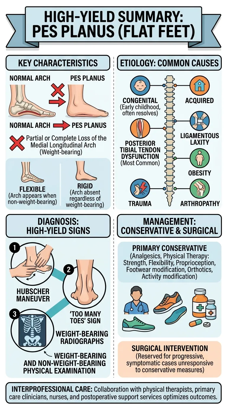
Pes planus, or flat feet, is a common foot deformity characterized by the partial or complete loss of the medial longitudinal arch during weight-bearing [1]. It can be either flexible, where an arch appears when non-weight-bearing, or rigid, where the arch is absent regardless of weight-bearing [1].
- Etiology: Can be congenital (common in early childhood, often resolves) or acquired (most commonly due to posterior tibial tendon dysfunction, ligamentous laxity, obesity, trauma, or arthropathy) [1].
- Clinical Impact: Ranges from an incidental finding to activity-limiting pain, altered gait, and progressive deformity affecting proximal joints [1].
- Diagnosis: Involves focused history, weight-bearing and non-weight-bearing physical examination (e.g., Hubscher maneuver, 'too many toes' sign, calcaneal valgus), and weight-bearing radiographs. Advanced imaging like MRI may assess soft tissues or specific osseous pathology [1].
- Management: Primarily conservative, including footwear modification, orthotics, activity modification, analgesics, and physical therapy focused on strength, flexibility, and proprioception. Surgical intervention is reserved for progressive, symptomatic cases unresponsive to conservative measures [1].
- Interprofessional Care: Collaboration with physical therapists, primary care clinicians, nurses, and postoperative support services optimizes outcomes [1].
Definition / Overview
Pes planus, commonly known as flat feet, is a foot deformity characterized by the partial or complete collapse or loss of the medial longitudinal arch, which contacts or nearly contacts the ground during weight-bearing [1]. This condition can be broadly categorized into two main types:
- Flexible Pes Planus: An arch is visible when the foot is not bearing weight but disappears upon standing [1]. This is the most common type, especially in children [1].
- Rigid Pes Planus: The arch is absent even when the foot is not bearing weight [1]. This form is less common and often indicative of underlying pathology [1].
The medial longitudinal arch is crucial for dissipating weight-bearing forces during the gait cycle [1]. While often asymptomatic, pes planus can alter lower extremity and lumbar spine biomechanics, increasing the risk of pain and injury to the foot, ankle, knees, and hips, sometimes necessitating intervention [1]. Pes planus can be either congenital or acquired [1].
Epidemiology
Pes planus is a prevalent condition, estimated to affect approximately 20% to 37% of the general population [1]. The majority of these cases are flexible pes planus [1]. Key epidemiological insights include:
- Age: More common in children, with most developing a normal arch by 10 years of age [1]. However, some children retain flat feet into adulthood [1].
- Sex: While some studies report a 3-fold higher prevalence among females, a 2023 review suggested a higher incidence among men in certain age groups [1].
- Ethnicity: A 2003 study found a prevalence of 17% among non-Hispanic White individuals and 34% among Black individuals [1]. A more recent review indicated a lower incidence in White populations and higher in Asian individuals [1].
- Weight: Overweight or obese children and adults show a higher prevalence of pes planus [1].
- Genetics: There is a strong genetic and familial predisposition to pes planus [1].
- Incidental Finding: The presence of pes planus, either radiographically or clinically, can be an incidental finding that requires clinical correlation to assess its significance [1].
Pathophysiology
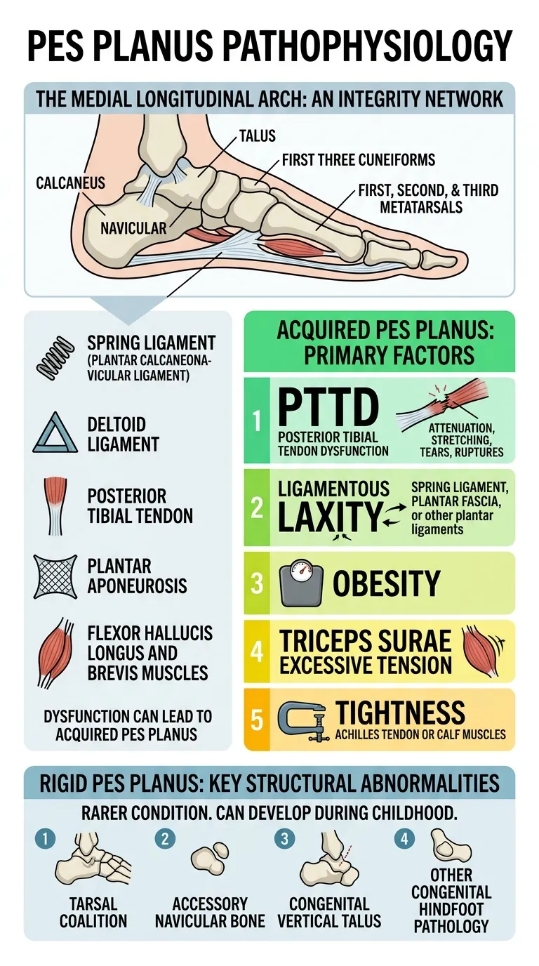
The medial longitudinal arch is a complex structure composed of the calcaneus, navicular, talus, the first three cuneiforms, and the first, second, and third metatarsals [1]. Its integrity is maintained by a network of soft tissue structures:
- Spring ligament (plantar calcaneonavicular ligament) [1]
- Deltoid ligament [1]
- Posterior tibial tendon [1]
- Plantar aponeurosis [1]
- Flexor hallucis longus and brevis muscles [1]
Dysfunction in any of these supporting elements can lead to acquired pes planus [1]. The primary factors contributing to acquired flatfoot deformity include:
- Excessive tension in the triceps surae [1]
- Obesity [1]
- Posterior tibial tendon dysfunction (e.g., attenuation, stretching, tears, ruptures) [1]
- Ligamentous laxity affecting the spring ligament, plantar fascia, or other plantar ligaments [1]
- Tightness of the Achilles tendon or calf muscles [1]
Rigid pes planus is a rarer condition that typically develops during childhood, though it can manifest at any age [1]. Its causes often involve structural abnormalities, such as:
- Tarsal coalition [1]
- Accessory navicular bone [1]
- Congenital vertical talus [1]
- Other forms of congenital hindfoot pathology [1]
Clinical Presentation
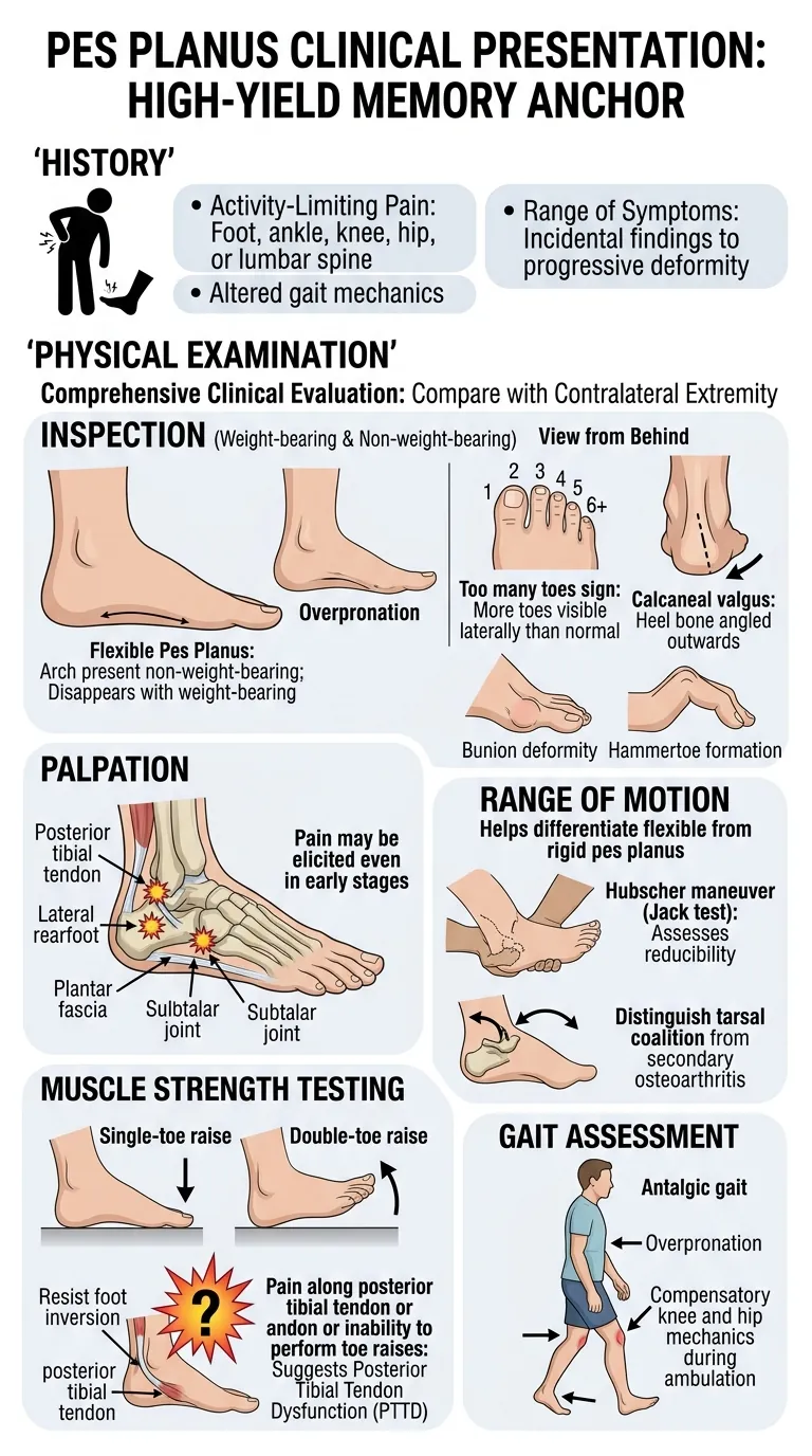
A comprehensive clinical evaluation for pes planus involves a focused history and a detailed physical examination, comparing findings with the contralateral extremity [1].
History
- Patients may report activity-limiting pain in the foot, ankle, knee, hip, or lumbar spine [1].
- Altered gait mechanics may be observed or reported [1].
- Symptoms can range from incidental findings to progressive deformity [1].
Physical Examination
The physical examination should include inspection, palpation, range of motion, muscle strength testing, and gait assessment [1].
Inspection
- Evaluate the patient in both weight-bearing and non-weight-bearing positions [1].
- In flexible pes planus, an arch is present non-weight-bearing but disappears with weight-bearing [1].
- Observe for overpronation [1].
- From behind, look for the 'too many toes' sign (more toes visible laterally than normal) and calcaneal valgus (heel bone angled outwards) [1].
- Compare both feet for asymmetry [1].
- Patients may also present with a bunion deformity or hammertoe formation [1].
Palpation
- Palpate the posterior tibial tendon, lateral rearfoot, and plantar fascia [1].
- Assess the subtalar joint [1].
- Pain may be elicited in these areas, even in early stages [1].
Range of Motion
- Helps differentiate flexible from rigid pes planus [1].
- The Hubscher maneuver (Jack test) assesses reducibility of the deformity [1].
- Subtalar joint range of motion testing can help distinguish tarsal coalition from secondary osteoarthritis [1].
- Pain or restricted motion may warrant further diagnostic studies [1].
Muscle Strength Testing
- Assess by having the patient perform single- and double-toe raises [1].
- Posterior tibial tendon strength is evaluated by resisting foot inversion [1].
- Pain along the posterior tibial tendon or inability to perform toe raises suggests posterior tibial tendon dysfunction [1].
Gait Assessment
- Patients may exhibit an antalgic gait [1].
- Observe for overpronation and compensatory knee and hip mechanics during ambulation [1].
Red Flags
⚠️ Do Not Miss: Rigid pes planus is a significant red flag, often indicating underlying structural pathology rather than a benign flexible flatfoot [1]. Conditions like tarsal coalition, accessory navicular bone, congenital vertical talus, or other congenital hindfoot pathologies can cause rigid pes planus and require specific management [1]. Persistent pain, restricted range of motion, or progressive deformity, especially in the presence of confirmed or suspected pes planus, also warrant prompt and thorough diagnostic evaluation [1].
Other red flags or concerns that point to specific etiologies and require advanced evaluation include:
- Pain or restricted motion during range of motion testing [1].
- Inability to perform a single toe raise or pain along the posterior tibial tendon, suggesting posterior tibial tendon dysfunction [1].
- Evidence of Charcot arthropathy in patients with sensory neuropathy, leading to midfoot collapse [1].
- Patients with arthropathies (e.g., rheumatoid arthritis, seronegative arthropathies) and poorly controlled disease are at higher risk for progressive acquired pes planus [1].
Diagnosis
The diagnostic approach for pes planus integrates a focused history and physical examination with imaging studies [1].
Plain Radiographs
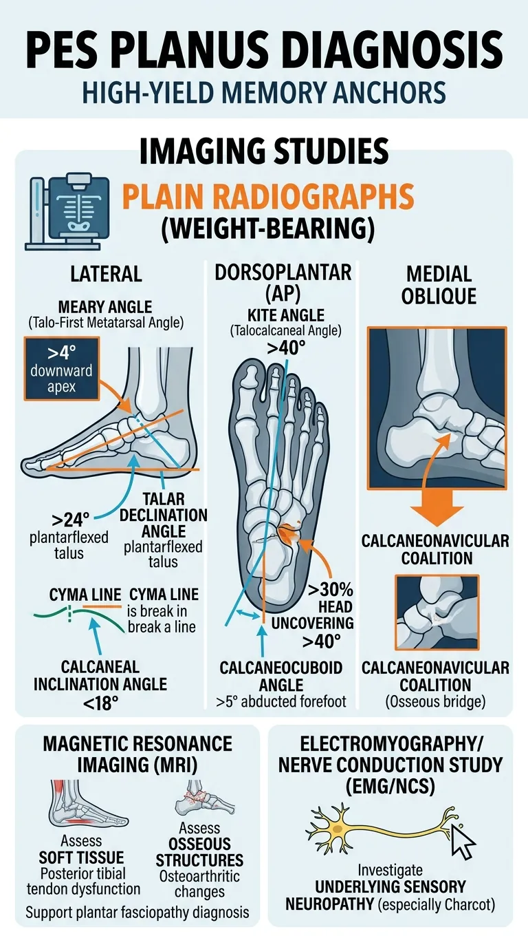
Three standard weight-bearing radiographs of the feet are typically sufficient: lateral, dorsoplantar (anteroposterior), and medial oblique views [1]. Additional views like a calcaneal axial may be obtained as needed [1].
Lateral Weight-Bearing Radiographs
- Meary Angle (Talo-First Metatarsal Angle): Normal is ~0°; >4° downward apex indicates longitudinal arch collapse at the talonavicular joint [1].
- Talar Declination Angle: Measures talus tilt; >24° suggests plantarflexed talus in flat foot [1].
- Calcaneal Inclination Angle: <18° indicates pes planus [1].
- Cyma Line: A break in this S-shaped line (between talonavicular and calcaneocuboid joints) suggests pes planus [1].
Medial Oblique Weight-Bearing Radiographs
- May reveal a calcaneonavicular coalition, indicated by close approximation or an osseous bridge between the anterior process of the calcaneus and the lateral navicular [1].
Dorsoplantar (Anteroposterior) Weight-Bearing Radiographs
- Calcaneocuboid Angle: >5° indicates an abducted forefoot on the rearfoot [1].
- Talar Head Uncovering: >30% uncovering of the talar head by the navicular articulation suggests medial talar head rotation [1].
- Kite Angle (Talocalcaneal Angle): Normal range 25°-40°; >40° indicates pes planus [1].
Magnetic Resonance Imaging (MRI)
MRI is valuable preoperatively for assessing soft-tissue and osseous structures [1].
- Soft Tissue: Identifies posterior tibial tendon dysfunction, or injury to the spring ligament or other supporting soft tissues [1].
- Osseous Structures: Reveals osteoarthritic changes, avascular necrosis, cystic changes, and tarsal coalition [1].
- May support plantar fasciopathy diagnosis in cases of heel pain (edema at plantar fascial calcaneal enthesis) [1].
Electromyography/Nerve Conduction Study (EMG/NCS)
- Can be performed to investigate underlying sensory neuropathy, especially in suspected Charcot arthropathy [1].
Management
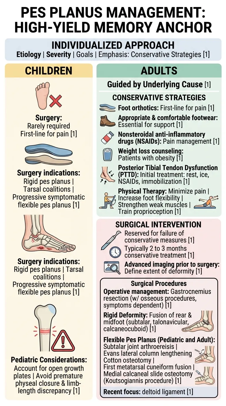
Management of pes planus is individualized based on etiology, symptom severity, and functional goals, with a strong emphasis on conservative strategies [1].
Children
- Surgical intervention is rarely required [1].
- Foot orthotics are often the first-line treatment for foot pain, especially when combined with leg, knee, or back pain [1].
- Surgery may be indicated for rigid pes planus, tarsal coalitions, or progressive, symptomatic flexible pes planus [1].
Adults
Treatment is guided by the underlying cause [1].
Conservative Strategies
- Foot orthotics: First-line therapy for pain [1].
- Appropriate and comfortable footwear: Essential for support [1].
- Nonsteroidal anti-inflammatory drugs (NSAIDs): For pain management [1].
- Weight loss: Counseling for patients with obesity is crucial [1].
- Posterior Tibial Tendon Dysfunction (PTTD): Initial treatment includes rest, ice, NSAIDs, and immobilization [1].
- Physical Therapy: Aims to minimize pain, increase foot flexibility, strengthen weak muscles, and train proprioception [1].
Surgical Intervention
Surgical procedures are reserved for patients who do not improve with conservative measures, typically after 2 to 3 months of conservative treatment [1]. More advanced imaging may be necessary prior to surgery to define the extent of deformity [1].
Surgical Procedures
Operative management may include gastrocnemius resection in conjunction with osseous procedures, depending on symptoms and deformity severity [1].
- Rigid Deformity: Fusion of the rear and midfoot is common, including subtalar, talonavicular, and calcaneocuboid fusions [1].
- Pediatric Considerations: Procedures must account for open growth plates to avoid premature physeal closure and limb-length discrepancy [1].
- Flexible Pes Planus (Pediatric and Adult): Procedures include subtalar joint arthroereisis, Evans lateral column lengthening, Cotton osteotomy, first metatarsal cuneiform fusion, and medial calcaneal slide osteotomy (Koutsogiannis procedure) [1].
- Recent focus also includes deltoid ligament complex repair and other soft-tissue adjunct procedures [1].
Postoperative and Rehabilitation Care
- Most patients use a protective non-weight-bearing brace for 6 to 8 weeks post-surgery [1].
- Total healing time varies and may be prolonged by physical therapy, overall health, and age [1].
- Custom orthotics are often required long-term to support surgical outcomes [1].
Complications
Complications associated with pes planus can arise from the condition itself or from surgical interventions [1].
Disease-Related Complications
- Progressive Deformity: Unmanaged or severe pes planus can lead to progressive deformity affecting the ankle, knee, hip, and lumbar spine [1].
- Pain and Injury: Increased risk of pain and injury to the foot, ankle, knees, and hips due to altered biomechanics [1].
- Specific Etiologies: Degenerative and inflammatory arthritis, as well as Charcot arthropathy, are often challenging to treat and are associated with progressive symptom worsening over time [1].
- Advanced Deformities: Associated with higher postoperative failure rates [1].
Postoperative Complications
Postoperative complications are dependent on the specific surgical procedure performed and can include [1]:
- Surgical site infection [1]
- Malunion/nonunion [1]
- Persistent pain [1]
- Implant dislocation/fracture [1]
- Other hardware complications [1]
- Recurrence of the deformity [1]
- Progressive arthritis [1]
Differential Diagnosis
While pes planus itself has various primary and secondary causes, it is important to distinguish it from conditions that may mimic its presentation or contribute to its development. The differential diagnosis, often clarified by clinical evaluation and imaging, typically includes [1]:
- Posterior Tibial Tendonitis/Dysfunction: Often a significant contributing factor to pain associated with acquired pes planus [1].
- Tarsal Coalition: A congenital fusion between two or more tarsal bones, commonly leading to rigid flatfoot [1].
- Arthritis (Primary or Secondary): Degenerative or inflammatory conditions (e.g., rheumatoid arthritis, seronegative arthropathies) can cause acquired pes planus [1].
- Connective Tissue Disorders: Conditions like Marfan syndrome, Ehlers-Danlos syndrome, or Down syndrome can cause ligamentous laxity leading to pes planus [1].
- Charcot Arthropathy: A destructive process affecting joints, often due to sensory neuropathy (e.g., in diabetes mellitus), leading to midfoot collapse [1].
Exam Pearls
Clinical Pearl: Always examine the patient in both weight-bearing and non-weight-bearing positions to differentiate flexible from rigid pes planus. The presence of an arch when non-weight-bearing but its collapse during weight-bearing is characteristic of flexible flatfoot [1]. This distinction is critical for guiding management [1].
- 'Too Many Toes' Sign: Observing the patient from behind, if more toes are visible laterally than normal, it suggests hindfoot valgus and forefoot abduction often associated with pes planus [1].
- Calcaneal Valgus: The outward angling of the heel bone is another key finding on posterior inspection [1].
- Hubscher Maneuver (Jack Test): Dorsiflexion of the great toe can test the flexibility and reducibility of the arch [1]. A reducible arch indicates flexible pes planus [1].
- Single- and Double-Toe Raises: Inability to perform these or pain along the posterior tibial tendon can indicate posterior tibial tendon dysfunction, a common cause of acquired flatfoot [1].
Common Traps / Pitfalls
Common Exam Trap: Do not miss rigid pes planus. While flexible flatfoot is common and often benign, rigid flatfoot is rare and typically signals a more significant underlying pathology such as tarsal coalition or congenital vertical talus. Failure to recognize rigidity can delay appropriate diagnosis and intervention [1].
- Incidental Findings: Radiographic or clinical presence of pes planus may be an incidental finding. Always correlate findings with patient symptoms and functional limitations to determine the necessity of intervention [1]. Asymptomatic pes planus often does not require treatment [1].
- Ignoring Proximal Symptoms: Pes planus can alter biomechanics throughout the lower extremities and lumbar spine, leading to pain in the ankle, knee, hip, and lower back. Do not solely focus on foot pain, but consider the broader impact of the deformity [1].
- Skipping Weight-Bearing Imaging: Non-weight-bearing radiographs can obscure the true extent of arch collapse. Always obtain weight-bearing views for accurate assessment of deformity [1].
- Overlooking Soft Tissue Causes: While osseous deformities are important, posterior tibial tendon dysfunction, spring ligament injury, and plantar fascia issues are common soft tissue contributors to acquired pes planus that require specific assessment [1].
FAQ
Q: What is the difference between flexible and rigid pes planus?
A: Flexible pes planus describes a foot where the arch is present when not bearing weight but disappears upon standing or weight-bearing. In contrast, rigid pes planus means the arch is absent regardless of whether the foot is bearing weight [1]. Flexible pes planus is much more common, especially in children, and often resolves spontaneously [1].
Q: When should pes planus in a child be surgically treated?
A: Children rarely require surgical intervention for pes planus. Surgery is generally indicated only for rigid pes planus (often due to conditions like tarsal coalitions) or for progressive, symptomatic flexible pes planus that has not responded to conservative measures [1].
Q: What are the primary conservative treatments for adult pes planus?
A: First-line conservative treatments for adults include foot orthotics, appropriate and comfortable footwear, activity modification, and nonsteroidal anti-inflammatory drugs (NSAIDs) for pain. Weight loss is also important for obese patients. For posterior tibial tendon dysfunction, rest, ice, and immobilization are initial steps, followed by physical therapy [1].
Q: How is pes planus diagnosed using imaging?
A: Diagnosis primarily relies on weight-bearing plain radiographs (lateral, dorsoplantar, and medial oblique views) to assess various angles and lines (e.g., Meary angle, talar declination angle, calcaneal inclination angle, Cyma line, Kite angle, talar head uncovering, calcaneocuboid angle). MRI may be used preoperatively to evaluate soft tissue integrity and subtle osseous changes [1].
References
- [1] Pes Planus (academic): https://www.ncbi.nlm.nih.gov/books/NBK430802/